Figures & data
Figure 1. A mummified coniferous cone with cytoplasm preserved from the Miocene (15–20 Ma) at Clarkia, Idaho, US. Specimen number PB20715, deposited in the Palaeobotanical Collection, Nanjing Institute of Geology and Palaeontology, Nanjing, China. (a) The fossil cone still embedded in the sediments. Note the cone axis (CA) and bract-scale complexes attached (arrows). Bar = 2 mm. (b) A cross view of a bract with the SEM. Note the intact cells and their contents (arrows) in the tissue. Bar = 20 µm. (c) A view of a bract thin section with the SEM. Note the epidermal and other cells, cytoplasm (CP) filling up the lumina, possible resin duct (RD), the cleavages between cells and their cell walls, and the ruptured cuticle (arrow). Section is about 2 µm thick. Bar = 10 µm. (d) A view of a cross-section of a bract-scale with the light microscope. Note the dark-colored cytoplasm in contrast to the light-colored cell walls in this unstained section. Bar = 0.1mm. This Figure is published with the permission of Nanjing Institute of Geology and Palaeontology, Nanjing, China and is reproduced in colour in Molecular Membrane Biology online.
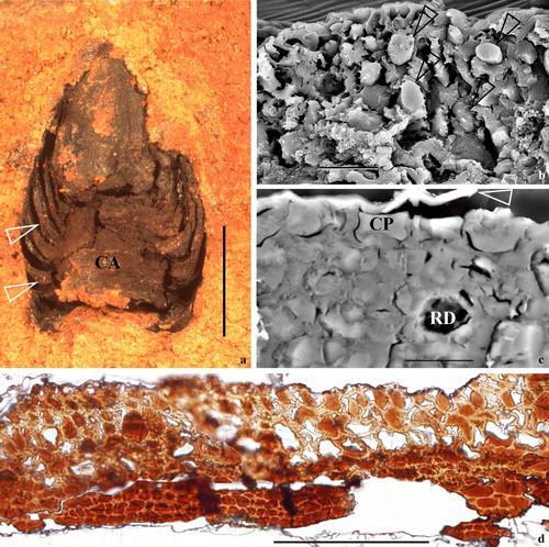
Figure 2. Vesicles and their relationships with CM in epidermal cells of a cone bract. All TEM electron micrographs. (a) A view of a cross-section of a bract. Note the faint-stained cell walls and dark-stained cytoplasm. The rectangular area is enlarged in b. Reproduced with permission from Wang (2007b). Bar = 5 µm. (b) A detailed view of an epidermal cell, enlarged from the rectangle in a. Note the dark-stained cytoplasm and faint-stained cell walls. The surface of the tissue is to the lower right. The rectangular region is shown in detail in c. Bar = 1 µm. (c) A detailed view of a portion of the cell shown in the rectangle in b. Note the relationship between the cuticle (CU), cell wall (CW), cytoplasmic membrane (CM), and cytoplasm (CP). Bar = 500 nm. (d) A detailed view of the vesicle fused to the CM, enlarged from the rectangle in c. Note that the vesicle's half-unit membrane is connected only with the inner leaflet of the CM (arrow), which becomes wavy near the fusion pore. The outer leaflet of the CM at the fusion pore is barely visible at the fusion pore. The CM is clearly two-layered. Reproduced with permission from Wang (Citation2007b). Bar = 100 nm. (e) A spherical vesicle with a half-unit membrane in the cytoplasm. Bar = 100 nm. (f) A vesicle connected with the CM through a cylindrical connection. Note the connection between vesicle membrane and the CM (black arrow), and the still-visible transverse membrane (white arrow) at the bottom of the vesicle. Bar = 100 nm. (g) The half-unit membrane of a vesicle connected with the inner leaflet of the CM. Note the connection between vesicle membrane and the inner leaflet of the CM (white arrow). Bar = 100 nm.
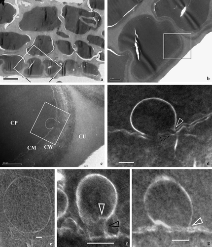
Figure 3. Vesicles’ membranes and their relationship with double-layered cytoplasmic membrane. All TEM electron micrographs. (a) A half-unit membrane of a vesicle connected to the inner leaflet of the CM. Note the connection between vesicle membrane and the inner leaflet of the CM (white arrow) in contrast to the dangling outer leaflet of CM (black arrow). Bar = 100 nm. (b) Half-unit membranes of several vesicles connected with the inner leaflet of the CM. Note the connection between vesicle membrane and the inner leaflet of the CM (white arrows) in contrast to the outer leaflet of CM (black arrow) parallel to the inner leaflet. Bar = 100 nm. (c) A half-unit membrane of another vesicle connected with the inner leaflet of the CM (white arrow). Note the dangling outer leaflet of CM (black arrow) and the drastic variation in CM thickness on each side of the vesicle. Bar = 100 nm. (d) Half-unit membranes of a vesicle connected with the CM. Note the connection between vesicle membrane and the inner leaflet of the CM (white arrows) in contrast to the dangling outer leaflet of CM (black arrow), and two more vesicles in the cell. Bar = 100 nm. (e) Several vesicles with half-unit membranes crowded along the CM. Note that the CM is highly disturbed with its double-layered structure sometimes still visible (white arrow), and that two adjacent vesicles may have their membranes closely spaced (black arrow). Bar = 100 nm. (f) A vesicle connected with the CM. The vesicle has a unit membrane (quite different from those in a–e, white arrow). The vesicle and the CM form an omega-shaped configuration with a narrow neck (black arrow). The vesicle membrane may become thicker and obscure due to oblique orientation of the membrane relative to the sectioning plane. Bar = 100 nm. (g) A typical double-layered structure of the CM. Bar = 100 nm. (h) A couple of the adjacent spherical vesicles before fusing with the CM. Bar = 200 nm. (i) Many vesicles with a half unit membrane in cytoplasm. Note the size and shape of the vesicles may vary from one to another. Bar = 200 nm. (j) More vesicles in the cytoplasm. Note the relationship between vesicles (arrows), and the varying membrane structure even within the same vesicle (lower arrow). Bar = 100 nm.
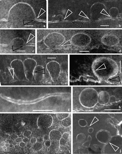
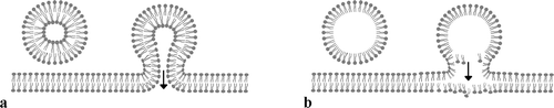
Figure 4. Diagram showing two different membrane fusion patterns. (a) A vesicle with a unit membrane and its fusion with double-layered cytoplasmic membrane. Note that both the inner and the outer leaflets of the CM are connected with their counterparts in the vesicle membrane. (b) A vesicle with a half-unit membrane and its fusion with double-layered CM. Note that only the inner leaflet of the CM is connected with the vesicle membrane while the outer leaflet of the CM is slightly distorted due to the impact of the vesicle content. This is a new pattern proposed based on the observation on cells in this fossil material.