
Figures & data
Figure 1. Emodin inhibits fibroblast fibrosis induced by ANGII. (a) CCK8 was used to examine changes in fibroblast proliferation after emodin treatment. (b) DHE was used to examine ROS levels in cardiac fibroblasts in each group, and red indicates positive results. (c) IPP was used to measure the mean density of the cells in each group. (d) IPP was used to measure the positive area of each group of cells. (e) Western blotting was used to examine changes in the expression of collagen II and α-SMA in each group, and GAPDH was used as the internal reference protein. (f) The relative gray values in each group. For each group of six mice, the data are expressed as the mean ± standard deviation (±S). **p < .01; *p < .05; ##p < .05.
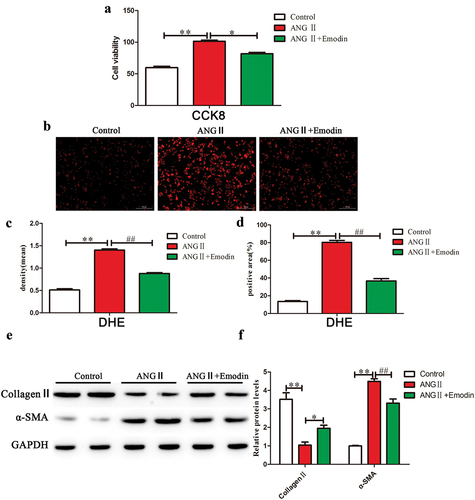
Figure 2. Emodin inhibits ANGII-induced cardiac fibrosis via the PI3K/AKT/mTOR signaling pathway (a,b) Western blotting was used to examine changes in the expression of PI3K/AKT/mTOR signaling pathway factors during cardiac fibrosis. GAPDH was used as an internal reference protein, and the gray value was calculated for p-AKT/AKT (c), p-mTOR/mTOR (d), α-SMA (e), and collagen II (F). **p < .01; ##p < .05; #p < .01.
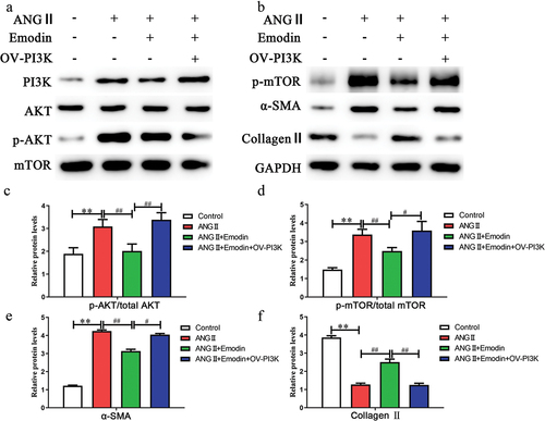
Figure 3. Emodin reduces oxidative stress in the heart and inhibits collagen fiber formation. (a) DHE was used to examine ROS levels in each group. (b) HE, Masson, echocardiography, electrocardiogram and Sirius red staining were used to examine changes in collagen fiber expression and cardiac function in each group. IPP was used to calculate the positive area (c) of each group. Changes in the heart mass index after emodin treatment in the MF group (d) and changes in collagen fiber formation (e,f). Changes in cardiac function (g,h). **p < .01; *p < .05.
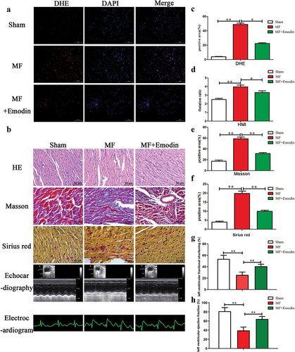
Figure 4. Emodin inhibits activation of the PI3K/AKT/mTOR signaling pathway and alleviates myocardial fibrosis. (a,b) Western blotting was used to examine changes in the expression of PI3K/AKT/mTOR signaling pathway components in the heart. GAPDH was used as an internal reference protein, and the gray value of p-AKT/AKT (c), p-mTOR/mTOR (d), α-SMA (e), and collagen II (f) was determined. **p < .01; ##p < .05.
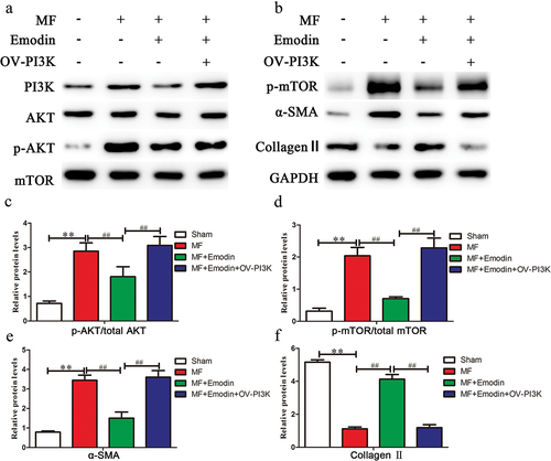
Figure 5. Emodin alleviates cardiac dysfunction after myocardial infarction. Echocardiography was used to evaluate changes in cardiac function, as indicated by LVEDD (a), LVESD (b), EF (c), FS (d), LVESP (e), LVEDP (f) and HR (g). **p < .01; *p < .05.
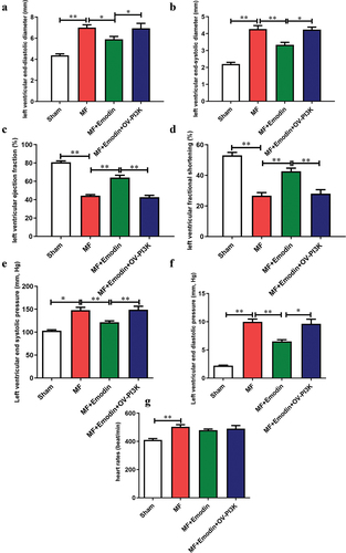
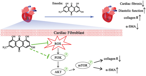
Figure 6. Simplified diagram showing the mechanism by which emodin alleviates MF.
Supplemental Material
Download MS Word (490.5 KB)Data availability statement
The data presented in this study are available upon request from the corresponding author.