971
Views
25
CrossRef citations to date
0
Altmetric
Research Article

Insulin-loaded poly(acrylic acid)-cysteine nanoparticles: Stability studies towards digestive enzymes of the intestine

, , &
Pages 254-260 | Received 02 Feb 2009, Accepted 26 Mar 2009, Published online: 30 Apr 2009

Figures & data

Figure 1. Particle size distributions directly after precipitation (-□-), after oxidation (-▴-), and after release from the lipid formulation (-⧫-). Indicated values are means ± SD (n ≥ 3).
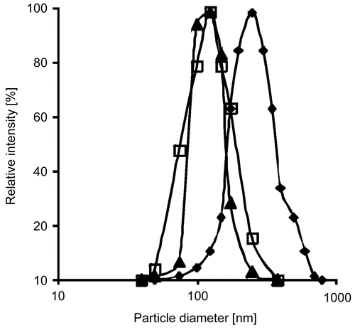
Figure 1.  Particle size distributions directly after precipitation (-□-), after oxidation (-▴-), and after release from the lipid formulation (-⧫-). Indicated values are means ± SD (n ≥ 3).

Table 1. Characterization of the utilized PAA100-Cys nanoparticles regarding mean particle diameter, zeta potential, and drug load. Indicated values are means ± SD (n ≥ 3).

Figure 2. In vitro release of insulin from PAA100-Cys nanoparticles as a percentage of the total amount of insulin present in the utilized formulation. Indicated values are means ± SD (n ≥ 3).
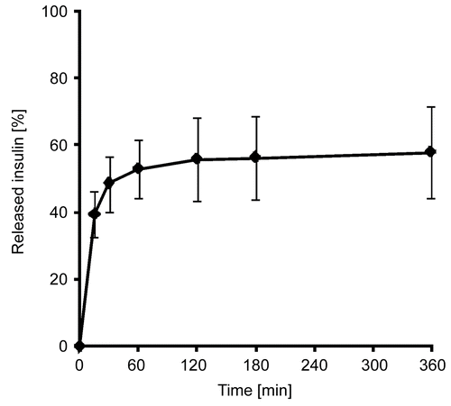
Figure 2.  In vitro release of insulin from PAA100-Cys nanoparticles as a percentage of the total amount of insulin present in the utilized formulation. Indicated values are means ± SD (n ≥ 3).

Figure 3. Degradation of insulin by trypsin in an aqueous solution (⧫), as a solid dispersion of insulin in triglyceride (▴), and as a solid dispersion of insulin-loaded PAA100-Cys nanoparticles in triglyceride (▪). Indicated values are means ± SD (n ≥ 3).
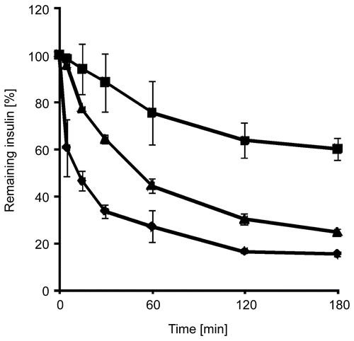
Figure 3.  Degradation of insulin by trypsin in an aqueous solution (⧫), as a solid dispersion of insulin in triglyceride (▴), and as a solid dispersion of insulin-loaded PAA100-Cys nanoparticles in triglyceride (▪). Indicated values are means ± SD (n ≥ 3).
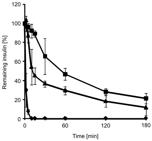
Figure 4. Degradation of insulin by α-chymotrypsin in an aqueous solution (⧫), as a solid dispersion of insulin in triglyceride (▴), and as a solid dispersion of insulin-loaded PAA100-Cys nanoparticles in triglyceride (▪). Indicated values are means ± SD (n ≥ 3).
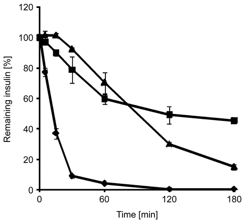
Figure 4.  Degradation of insulin by α-chymotrypsin in an aqueous solution (⧫), as a solid dispersion of insulin in triglyceride (▴), and as a solid dispersion of insulin-loaded PAA100-Cys nanoparticles in triglyceride (▪). Indicated values are means ± SD (n ≥ 3).

Figure 5. Degradation of insulin by elastase in an aqueous solution (⧫), as a solid dispersion of insulin in triglyceride (▴), and as a solid dispersion of insulin-loaded PAA100-Cys nanoparticles in triglyceride (▪). Indicated values are means ± SD (n ≥ 3).

Figure 5.  Degradation of insulin by elastase in an aqueous solution (⧫), as a solid dispersion of insulin in triglyceride (▴), and as a solid dispersion of insulin-loaded PAA100-Cys nanoparticles in triglyceride (▪). Indicated values are means ± SD (n ≥ 3).

Reprints and Corporate Permissions

Please note: Selecting permissions does not provide access to the full text of the article, please see our help page How do I view content?

To request a reprint or corporate permissions for this article, please click on the relevant link below:

Academic Permissions

Please note: Selecting permissions does not provide access to the full text of the article, please see our help page How do I view content?

Obtain permissions instantly via Rightslink by clicking on the button below:

If you are unable to obtain permissions via Rightslink, please complete and submit this Permissions form. For more information, please visit our Permissions help page.