Figures & data
Figure 1. Phage binding to Hepa 1c1c7 cells and MEF. (A and B) Hepa 1c1c7 cells incubated with liver-targeting phage 20-6F in OptiMEM in the presence or absence of Lf or RAP. (C) Wild-type and LRP-deficient MEF (congenic cell line PEA-13) incubated with liver-targeting phage 20-6F or non-targeting phage 20-6 in OptiMEM. The amount of cell-bound phage was measured by a plaque assay. Duplicate samples were used. Data are shown as mean ± SE.
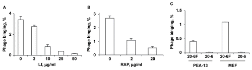
Figure 2. Reduced phage uptake by LRP-deficient liver in Ad5/Cre-injected mice. (A) Phage contents in liver and blood were determined by a plaque assay. Data are shown as mean ± SD, n = 3. (B) Immunohistochemistry of normal liver after phage injection. (C) Immunohistochemistry of LRP-deficient liver after phage injection. Red, green, and blue colors correspond to phage, actin, and nuclei, respectively.
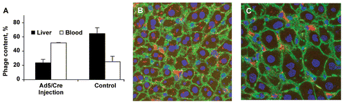
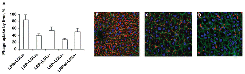
Figure 3. Reduced phage uptake by LRP-deficient liver in ‘floxed’ LRP mice expressing Cre. (A) Liver phage uptake as determined by a plaque assay. Data are shown as mean ± SD, n = 4. (B) Confocal images of immunostained LRP+LDLr+ liver sections (Flox+/+, Cre−, LDLr+/+). (C) Confocal images of immunostained LRP−LDLr+ liver sections (Flox+/+, Cre+, LDLr+/+). (D) Confocal images of immunostained LRP−LDLr+ liver sections (Flox+/+, Cre+, LDLr−/−). Red, green, and blue image colors correspond to phage, actin, and nuclei, respectively.