3,930
Views
20
CrossRef citations to date
0
Altmetric
Research Articles

Hydrogels for localized chemotherapy of liver cancer: a possible strategy for improved and safe liver cancer treatment

, , , , , & ORCID Icon show all
Pages 1457-1476 | Received 07 Feb 2022, Accepted 11 Apr 2022, Published online: 09 May 2022

Figures & data

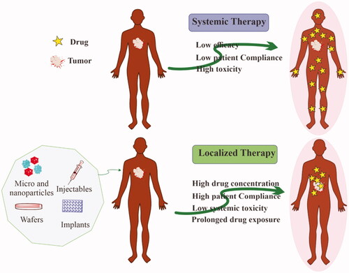
Figure 1. Advantages of localized chemotherapy over conventional systemic chemotherapy for the treatment of solid tumors.

Figure 1. Advantages of localized chemotherapy over conventional systemic chemotherapy for the treatment of solid tumors.

Table 1. Hydrogel-based drug delivery systems for cancer treatment.

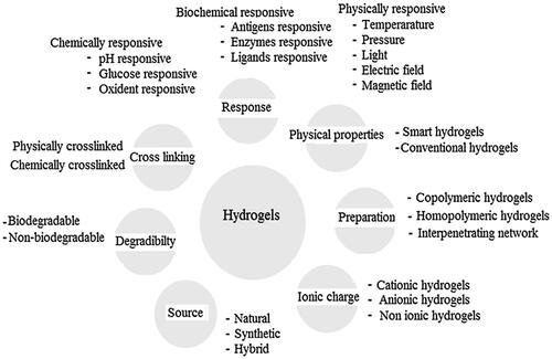
Figure 2. Classification of hydrogels based on the different properties. Reproduced with permission from reference Ullah et al. (Citation2015).

Figure 2. Classification of hydrogels based on the different properties. Reproduced with permission from reference Ullah et al. (Citation2015).

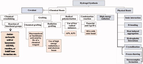
Figure 3. Various physical and chemical approaches for the synthesis of the hydrogel. Reproduced with permission from reference Sinha & Chakma (Citation2019).

Figure 3. Various physical and chemical approaches for the synthesis of the hydrogel. Reproduced with permission from reference Sinha & Chakma (Citation2019).

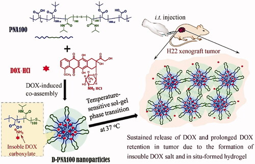
Figure 4. D-PNAx hydrogels for i.t. injection of targeted chemotherapy using DOX-induced co-assembling nanoparticles are shown schematically. Reproduced with permission from Wan et al. (Citation2016).

Figure 4. D-PNAx hydrogels for i.t. injection of targeted chemotherapy using DOX-induced co-assembling nanoparticles are shown schematically. Reproduced with permission from Wan et al. (Citation2016).

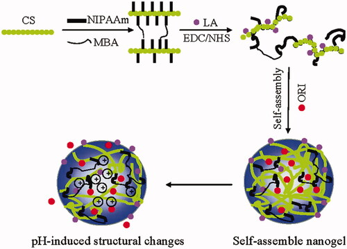
Figure 5. Schematic diagram depicting the concept of Gal-CS-g-PNIPAM nanogels. Reproduced with permission from American Chemical Society 2011 (Duan et al., Citation2011).

Figure 5. Schematic diagram depicting the concept of Gal-CS-g-PNIPAM nanogels. Reproduced with permission from American Chemical Society 2011 (Duan et al., Citation2011).

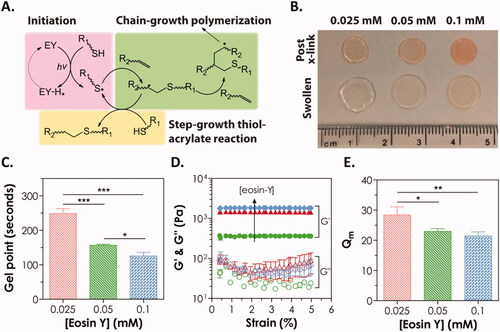
Figure 6. (A) Mechanism of visible light initiated mixed-mode step-chain-growth thiol-acrylate photopolymerization. (B–E) Physical properties of visible light-cured PEGDA thiol-acrylate hydrogels formed with 0.025, 0.05, or 0.1 mM of eosin-Y. (B) Photographs; (C) gel points; (D) elastic (G′) and viscous (G″) modulus, and (E) equilibrium mass swelling ratio (Qm). Reproduced with permission from Wiley Online Library (Hao & Lin, Citation2014).

Figure 6. (A) Mechanism of visible light initiated mixed-mode step-chain-growth thiol-acrylate photopolymerization. (B–E) Physical properties of visible light-cured PEGDA thiol-acrylate hydrogels formed with 0.025, 0.05, or 0.1 mM of eosin-Y. (B) Photographs; (C) gel points; (D) elastic (G′) and viscous (G″) modulus, and (E) equilibrium mass swelling ratio (Qm). Reproduced with permission from Wiley Online Library (Hao & Lin, Citation2014).

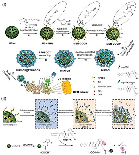
Figure 7. (I) Schematic illustration of MSN-GII@PFH&DOX includes (a) the MSNs surface modification with amino groups and (b, c) carboxyl groups activation, (d) in situ amidation-initiated self-assembled supramolecular nanogels on the interface of MSNs (MSN-GI), (e) ternary inorganic-supramolecular-polymeric nanogels (MSN-GII) by laccase-mediated polymerization, (3) co-loading of guest molecules PFH and DOX, and (4) ultrasound imaging and imaging-guided HIFU ablation of tumors. (II) Schematic illustration of the mechanism of in situ amidation-fueled self-assembly. Reproduced with permission from American Chemical Society 2019 (Wang et al., Citation2019).

Figure 7. (I) Schematic illustration of MSN-GII@PFH&DOX includes (a) the MSNs surface modification with amino groups and (b, c) carboxyl groups activation, (d) in situ amidation-initiated self-assembled supramolecular nanogels on the interface of MSNs (MSN-GI), (e) ternary inorganic-supramolecular-polymeric nanogels (MSN-GII) by laccase-mediated polymerization, (3) co-loading of guest molecules PFH and DOX, and (4) ultrasound imaging and imaging-guided HIFU ablation of tumors. (II) Schematic illustration of the mechanism of in situ amidation-fueled self-assembly. Reproduced with permission from American Chemical Society 2019 (Wang et al., Citation2019).

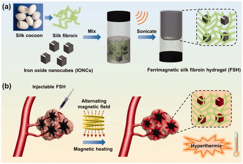
Figure 8. (a) Schematic illustration of the preparation of ferrimagnetic silk fibroin hydrogel (FSH); (b) injectable FSH guided magnetic hyperthermia therapy of tumor under alternating magnetic field (AMF). Reproduced with permission from reference (Qian et al., Citation2020).

Figure 8. (a) Schematic illustration of the preparation of ferrimagnetic silk fibroin hydrogel (FSH); (b) injectable FSH guided magnetic hyperthermia therapy of tumor under alternating magnetic field (AMF). Reproduced with permission from reference (Qian et al., Citation2020).

Table 2. List of stimuli-responsive hydrogel for liver cancer therapy.

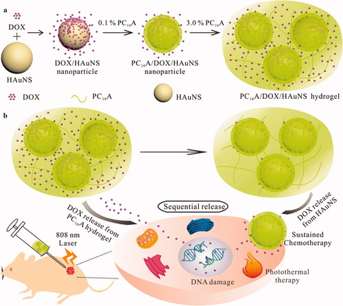
Figure 9. (a) In this schematic, we show the steps involved in developing PC10A/DOX/HAuNS hydrogels and nanoparticles, as well as the steps involved in dissolving the nanoparticles in the PC10A hydrogel. (b) The sequential drug release of PC10A/DOX/HAuNS hydrogels was used for long-term chemotherapy and photothermal therapy in vivo (Jin et al., Citation2019).

Figure 9. (a) In this schematic, we show the steps involved in developing PC10A/DOX/HAuNS hydrogels and nanoparticles, as well as the steps involved in dissolving the nanoparticles in the PC10A hydrogel. (b) The sequential drug release of PC10A/DOX/HAuNS hydrogels was used for long-term chemotherapy and photothermal therapy in vivo (Jin et al., Citation2019).

Table 3. Hydrogel-based active targeting and combination therapy for liver cancer therapy.

Table 4. Advantages and shortcomings of different hydrogel delivery routes for cancer treatment.