Figures & data
Figure 1. Location of intrathecal catheter tip confirmed by the fluoroscopy during surgery. The red arrow indicates the intrathecal catheter; the white arrow indicates the tip of catheter; white dash cycle indicates the foramen magnum; red dash cycle indicates the clivus; the yellow dash cycle indicates the pituitary fossa.
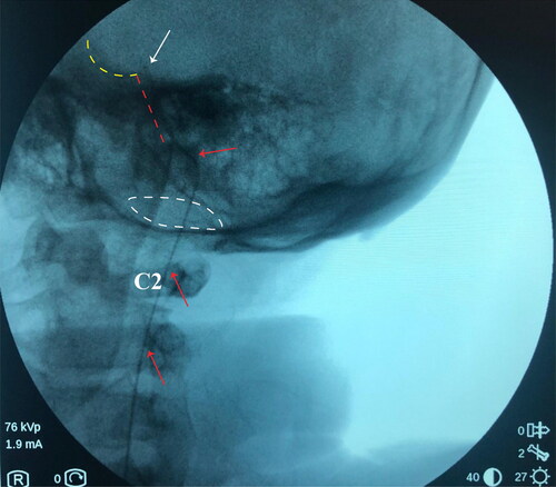
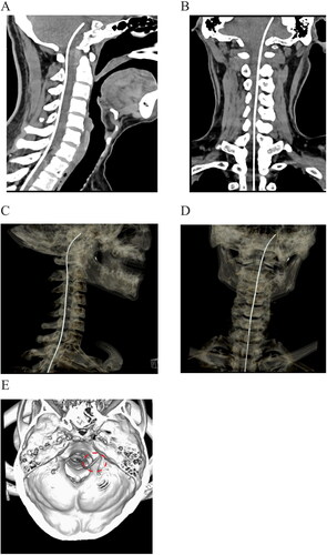
Figure 2. Postoperative imaging of catheter placement with computed tomography. a, b: Sagittal and coronal plane of scanning demonstrated that the tip of intrathecal catheter was located at the level of posterior clinoid process. c, d: Three-dimensional reconstruction of catheter placement by computed tomography. e: Reconstruction of the skull base to reveal the location of the catheter (circled by the red dash line).