Figures & data
Figure 1. Short-term stability for plasma samples at pH 7.4 at different temperatures. Treosulfan stability in plasma was evaluated at different temperature conditions (room temperature, +4 °C and −20 °C) for short-term incubation at pH 7.4 over several time points up to 8 h and different treosulfan concentrations. Treosulfan degraded almost in the same pattern at different temperatures regardless the drug concentration. At room temperature, treosulfan was stable up to 80% in the first 2 h, after that it started to be metabolized. The rate of treosulfan degradation at +4 °C was slower than what was obtained at room temperature. Frozen samples at −20 °C had the highest stability; treosulfan stability was 91% at 8 h. Plasma samples containing treosulfan can be safely stored at −20 °C for short period up to 8 h.
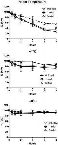
Table 1. Treosulfan stability in plasma at different conditions.
Table 2. Treosulfan stability in urine at different conditions.
Table 3. Treosulfan stability in cell culture media at different conditions.
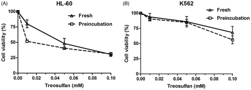
Figure 2. Cytotoxicity of treosulfan on (A) HL-60 and (B) K562 cells following incubationfor 24 h at 37 °C. The cytotoxicity of treosulfan on the leukemic cells HL-60 and K562 was determined through the WST-1 assay. The HL-60 cell line was more sensitive to treosulfan compared with K562 cell line. The HL-60 cells decrease in cell viability was concentration dependent. On the other hand, no toxicity was found in the K562 cells up to a concentration of 0.1 mM treosulfan. The IC50 of the HL-60 cells was 9.2 μM after 24 h and 1.6 μM after 48 h. In K562 cells, the IC50 was 151 μM after 24 h and 62 μM after 48 h treatment. Treosulfan cytotoxicity increased following pre-incubation for 24 h at 37 °C. Only 52% of the HL-60 cells were viable after treatment with the pre-incubated 0.01 mM treosulfan compared to 80% of HL-60 cells following treatment with freshly prepared treosulfan at the same concentration (A). On the other hand; 90% of the K562 cells were viable following incubation with 0.01 mM treosulfan with or without pre-incubation (B).