Figures & data
Figure 1 Ash and ammonia contents of pidan white during pickling and aging in the presence or absence of different cations. Bars represent the standard deviations (n = 3). Different letters on the bars within the same pickling/aging time indicate significant differences (P < 0.05).
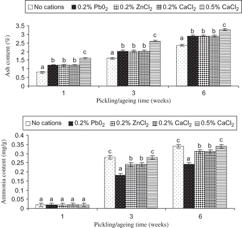
Figure 2 Ash and ammonia contents of pidan yolk during pickling and aging in the presence or absence of different cations. Bars represent the standard deviations (n = 3). Different letters on the bars within the same pickling/aging time indicate significant differences (P < 0.05).
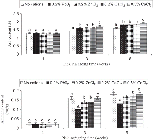
Figure 3 Changes in TCA-soluble peptides contents of pidan white treated without and with different cations during pickling and aging. Bars represent the standard deviations (n = 3). Different letters on the bars within the same pickling/aging time indicate significant differences (P < 0.05).
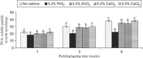
Figure 4 Scanning electron microscopic photograph of pidan white after three weeks of pickling. Magnification: 5X.
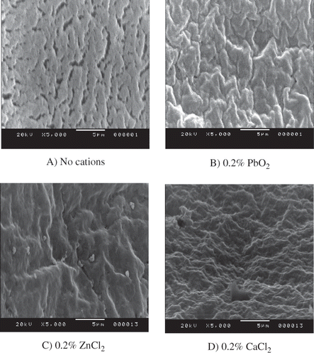
Figure 5 Confocal laser scanning microscope (CLSM) micrographs of exterior yolk for (3A)protein distribution, (3B) lipid distribution, and (3C) and combined image of protein and lipid of pidan treated with different cations after pickling (week 3). Pb: 0.2% PbO2; Zn: 0.2% ZnCl2; Ca: 0.2% CaCl2. Magnification: 200X (zoom X2.5).
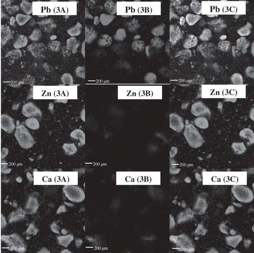
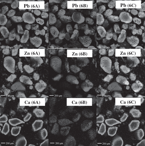
Figure 6 Confocal laser scanning microscope (CLSM) micrographs of exterior yolk for protein distribution (6A), lipid distribution (6B) and combined image of protein and lipid (6C) of pidan with different treatments after aging (week 6). Pb: 0.2% PbO2; Zn: 0.2% ZnCl2; Ca: 0.2% CaCl2. Magnification: 200X (zoom X2.5).