Figures & data
Figure 1 PAGE of extracted native myosin.
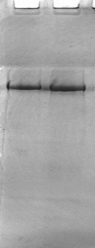
Table 1 Effect of setting time on surimi gel properties
Figure 2 Variations of chemical interactions during the formation of suwari gels and surimi gels. In horizontal axis, the numbers 1, 2, 3, 4, 5, 6, 7, 8, and 9 represent surimi, suwari gel setting at 40°C for 30 min, suwari gel setting at 40°C for 60 min, suwari gel setting at 40°C for 90 min, suwari gel setting at 40°C for 120 min, surimi gel setting at 40°C for 30 min, surimi gel setting at 40°C for 60 min, surimi gel setting at 40°C for 90 min, and surimi gel setting at 40°C for 120 min, respectively.
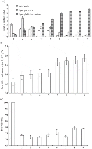
Figure 3 Laser Raman spectroscopy. A: surimi; B: suwari gel setting for 60 min at 40°C; C: surimi gel setting for 60 min at 40°C and cooking for 30 min at 90°C.
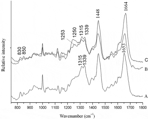
Table 2 Percent of secondary structure units of myosin
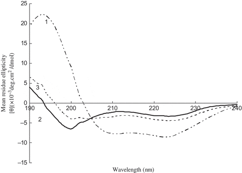
Figure 4 CD spectra of myosin during heating. The numbers 1, 2, and 3 represent native myosin (pH 7.5), myosin heating for 60 min at 40°C, myosin heating for 60 min at 40°C, and then heating for 30 min at 90°C, respectively.