Figures & data
Figure 1. SDS-PAGE patterns of proteins extracted from Bromelia pinguin L. fruit pulp. Lane 1: molecular marker; Lane 2: crude protein extract; Lane 3: soluble protein extract; Lane 4: membrane protein extract. A: Silver staining; B: Coomassie staining.
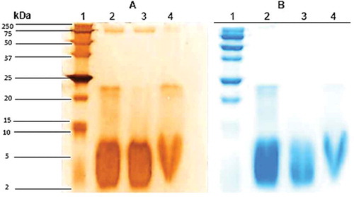
Figure 2. Chromatography elution profile of soluble protein extract of Bromelia pinguin fruit pulp, on fractionation using gel filtration chromatography.
Figure 3. Chromatograms obtained by reverse-phase high-performance liquid chromatography. A: Profile of soluble protein extract (SPE); B: Profile of peak one (PI).
Table 1. Extension of zone of bacteria growth inhibition around filter papers with samples in mm.
Figure 4. Time kill studies of E. coli ATCC 25922. A: and S. aureus ATCC 25923; B: exposed to ampicillin (AMP); tetracycline (TET); rifampicin (RIF); vancomycin (VAN); and Peak one (P1).
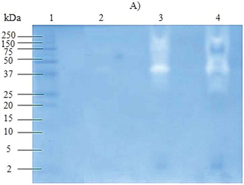
Figure 5. SDS-zymograph of proteases extracted from Bromelia pinguin L. fruit pulp. Lane 1: molecular marker; Lane 2: soluble protein extract; Lane 3: Peak 1; Lane 4: Peak 2.