Figures & data
Table 1. Antibacterial activity of POFE from P. oleracea.
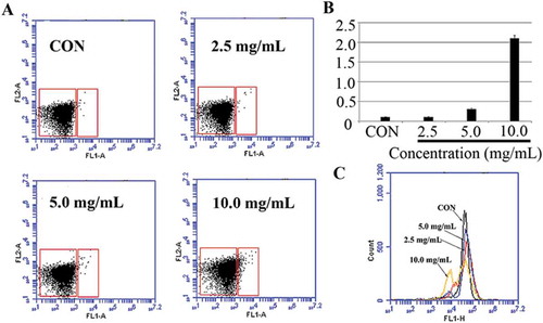
Figure 1. POFE-induce S. aureus intracellular ROS accumulation. S. aureus cells untreated with POFCE were as the control.
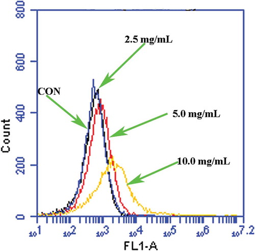
Figure 2. POFE-induce S. aureus cells apoptosis-like response. (A) and (B) Results of Annexin V/PI staining. Cells were sorted into living, necrotic, early apoptotic, and late apoptotic cells. (C) and (D). Hoechst 33342/PI double staining, Cells were sorted into living, apoptotic and dead cells. (E) Results of TUNEL assays. TUNEL positive cells (green channel). Hoechst 33342 (blue channel) is used to locate the nuclei of the cells. (F) Flow cytometric analysis of caspase-like response using FITC-VAS-FMK. S. aureus cells untreated with POFE were as the control.
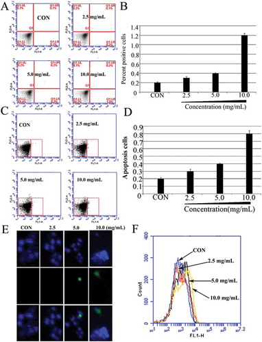
Figure 3. Membrane depolarisation analysis in POFE-treated S. aureus cells. (A) and (B) Mitochondrial Membrane Potential/Annexin V Apoptosis Kit analysed apoptosis based on PS translocation and changes in mitochondrial membrane potential. (C) Membrane potential (??m) was determined by using Rhodamine 123 staining and analysed by using flow cytometry. S. aureus cells untreated with POFE were as the control.