
Figures & data
Figure 1. Hardness of myofibrillar protein gel heated at different temperatures
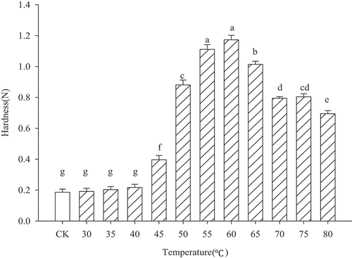
Figure 2. Water-holding capacity of myofibrillar protein gel heated at different temperatures
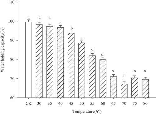
Figure 3. Reactive and total sulfhydryl (SH) groups of myofibrillar protein gel heated at different temperatures
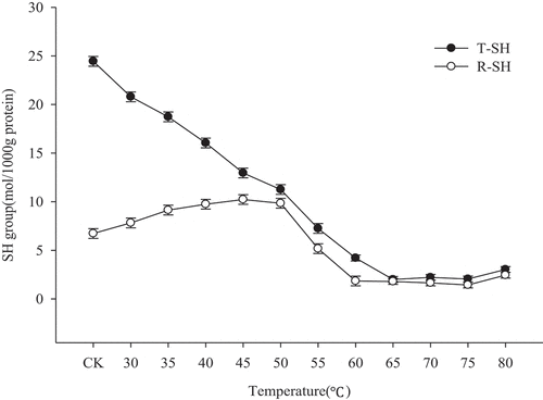
Figure 4. SDS-PAGE of supernatants protein in myofibrillar protein gel heated at different temperatures
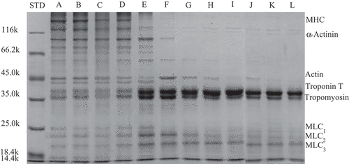
Figure 5. Raman spectra in 400–2000 cm−1 region of myofibrillar protein gels heated at different temperatures
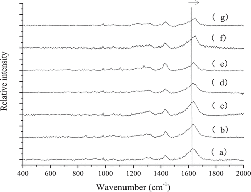
Figure 6. Raman spectra in 400–2000 cm−1 region of myofibrillar protein gels heated at different temperatures
Figure 7. S-S stretch in 450–600 cm−1 region of myofibrillar protein gel heated at different temperatures
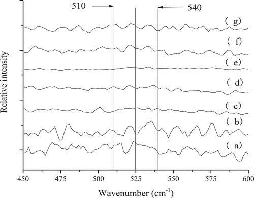
Table 1. Normalized intensity of the 760 cm−1 band, the 1450 cm−1 band, and normalized ratio of I850/I830 doublet bands
Table 2. Pearson correlation of gel properties and secondary structure of myofibrillar protein gels heated at different temperatures