Figures & data
Table 1. Real-time quantitative PCR primers used in this study
Figure 1. The effect of unhulled red djulis on cell cytotoxicity and wound healing. (A) CCD-966SK cells were treated with unhulled djulis extract (0.125%, 0.25%, 0.5%, 1%). Then, cytotoxicity was assessed through MTT assay. (B) Scrape with a sterile 1-ml pipette tip and then CCD-966SK cells were cultured with unhulled djulis extract (0.125%, 0.25%, 0.5%, 1%), and then using Image J software to analysis. (n = 3; mean ± SD, using a paired Student’s t-test, *p < .05 compared with control group)
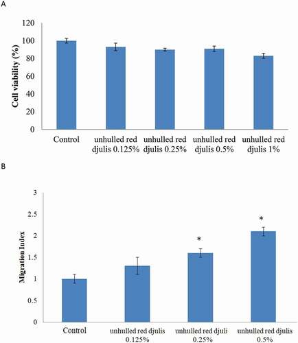
Figure 2. Unhulled red djulis extracts regulated TGM1, KRT1, KRT10, SOD2, COL1A2 and MMP9 expression. CCD-966SK cells were treated with unhulled djulis extract (0.5%), and then used qPCR analysis. (A) Skin related genes: TGM1, KRT1, KRT10, antioxidant-related genes: SOD2. (B) Collagen-related genes: COL1A2 and MMP9. (n = 3; mean ± SD, using a paired Student’s t-test, *p < .05 compared with control group)
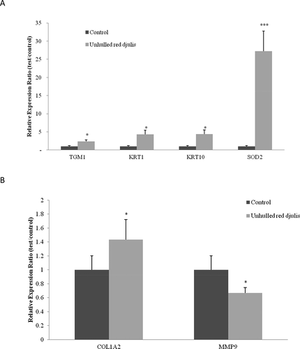
Figure 3. The effect of unhulled red djulis extracts on formation of AGEs and melanin. CCD-966SK cells were treated with unhulled djulis extract (0.5%), and then (A) analyzed the AGEs through advanced glycation end-products assay. (B) Analyzed the melanin through tyrosinase activity inhibition assay. (n = 3; mean ± SD, using a paired Student’s t-test, *p < .05 compared with control group)
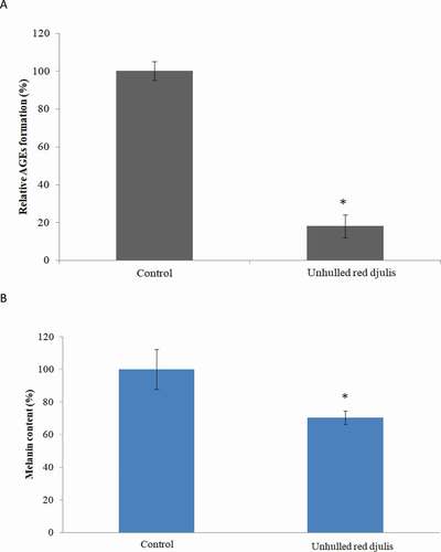
Figure 4. Effects of unhulled red djulis extracts on collagen content. CCD-966SK cells were treated with unhulled djulis extract (0.5%), and then analyzed the collagen by immunofluorescence staining. Green color: collagen. Blue color: Nucleus. (n = 3; mean ± SD, using a paired Student’s t-test, *p < .05 compared with control group)
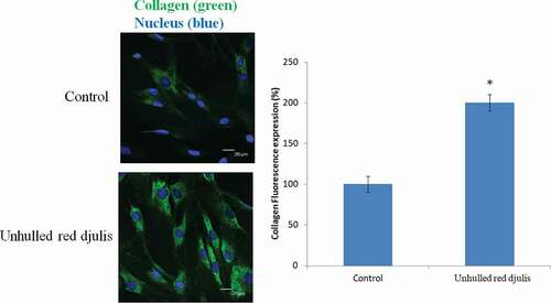
Table 2. Antioxidant capacity of djulis extract
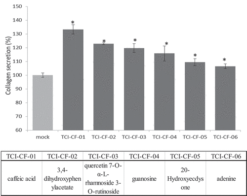
Figure 5. Effective components of unhulled red djulis extracts increased collagen content. (A) Profiles of unhulled red djulis extracts and compound structures. (B) CCD-966SK cells treated with 0.2 mg/mL of compounds 1–6, and then analyzed by collagen secretion assay. (n = 3; mean ± SD, using a paired Student’s t-test, *p < .05 compared with control group)