Figures & data
Figure 1. Total phenols and flavonoid content of R. sativus aqueous and methanol extracts. Values are expressed as means ± SD. Radish aqueous extract is richer on phenols and flavonoids than methanol extract. All experiments were made in triplicates.
Figure 2. HPLC profiles of the obtained compounds. Five phenolic compounds were identified in radish aqueous extract.
Table 1. Retention time (min) and concentration of phenolics identified in R. sativus bulb aqueous extract bu HPLC DAD.
Figure 3. GC-MS chromatogram of R. sativus methanol extract.
Figure 4. Chemical structure of 4-methylthio-3-butenyl isothiocyanate (raphasatin).
Table 2. IC50 of DPPH, ABTS, hydroxyl radical scavenging activities, ferric-reducing power and iron chelation of R. sativus bulb extracts.
Figure 5. Effect of R. sativus extracts on U-87 MG cells viability.. U-87 MG cells were incubated for 24, 48 and 72 h with different concentrations of R. sativus extracts (A) Methanol extract; (B) Aqueous extract. Doxorubicin (Dox) was used as positive control and water as negative one. All experiments were made in triplicates. Values were expressed as means ± SD, *Indicates significant differences (p < .05).
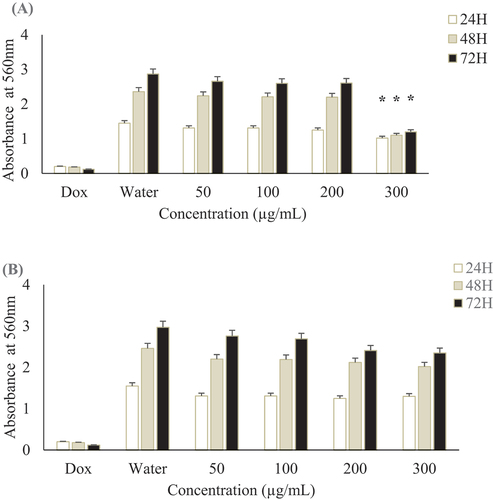
Figure 6. Radish methanol extract effect on various ECM in cell adhesion assays. Glioblastoma cells were preincubated with 10, 20, 50 and 100 µg/mL in radish methanol extract for 30 min at room temperature and then added to wells coated with 10 µg/mL fibronectin (Fn), 10 µg/mL fibrinogen (Fg) or 50 µg/mL (PLL) and allowed to attach for 1 h at 37°C. Values were expressed as means ± SD, *Indicates significant differences (p < .05).
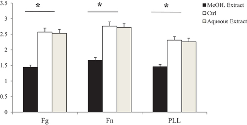
Figure 7. Cell adhesion assay of (A) 4-methylthio-3-butenyl isothiocyanate and (B) positive control doxorubicin (Dox) on different ECM proteins. 4-methylthio-3-butenyl isothiocyanate and posititive control doxorubicin inhibited with the same manner the adhesion of U-87 MG to fibronectin (Fn), fibrinogen (Fg) and poly-L-Lysine (PLL).
Table 3. IC50 (µg/mL) of radish extracts, raphasatin, and positive control doxorubicin against the tested ECM in dis-adhesion test.
Figure 8. 4-methylthio-3-butenyl isothiocyanate and doxorubicin (Dox) effects on U-87 MG cells proliferation. After 4 days of incubation with 0.5 µg/mL of raphasatin or doxorubicin, wells were washed with PBS and the cells were fixed with 1% glutaraldehyde, stained with 0.1% crystal violet and quantified by absorbance at 560 nm.