Figures & data
Figure 1. Construction of the quasi-spider silk protein gene. The monomeric spider silk gene (A); the triple helix-forming T blocks (B) at both ends of the triblock copolymers consisting of (Pro–Gly–Pro)15 homopolymeric stretches.
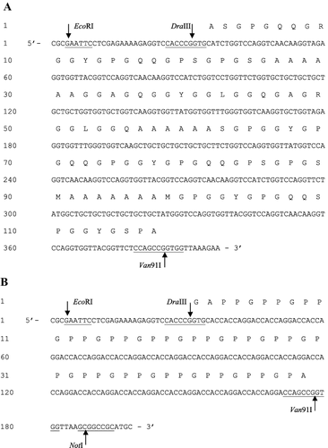
Figure 2. Construction of yeast expression vector pPIC9K-TS2T.
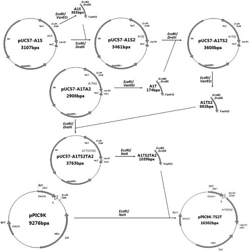
Figure 3. Identification of recombinant vector pPIC9K-TS2T by single enzyme digestion. Lane 1, non-linearized plasmid; Lane 2, linearized plasmid pPIC9K-TS2T (SacI); M, DL15000 DNA Maker.
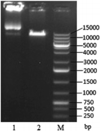
Figure 4. Identification of recombinant vector pPIC9K-TS2T by double enzyme digestion. Lane 1, linearized plasmid pPIC9K-TS2T (EcoRI/NotI); M, DL10000 DNA Maker.
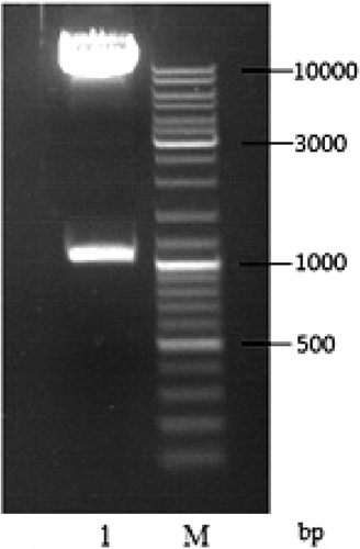
Figure 5. Colony PCR analysis of recombinant GS115 strains after electroporation. Lanes 1–12, GS115/pPIC9KTS2T; P, GS115/pPIC9K PCR product; M, DL15000 DNA Maker; N, GS115 PCR product.
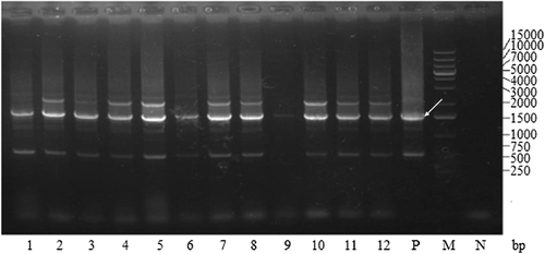
Figure 6. SDS-PAGE analysis of the quasi-spider silk protein expression and purification. Lane M, protein marker, Mid. Range; Lane 1, fermentation supernatant; Lane 2, purified sample.
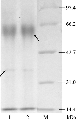
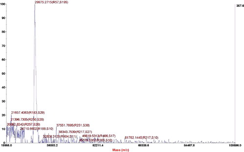
Figure 7. Mass-spectrographic analysis of quasi-spider silk protein.