
Figures & data
Figure 1. Study design: antenatal diagnosis and clinical outcome.
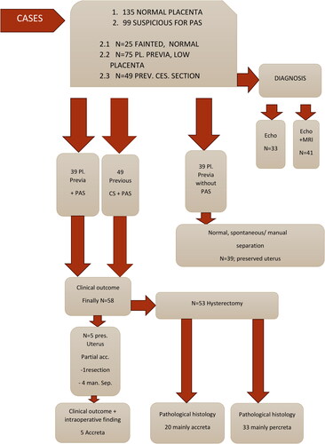
Figure 2. Normal appearance of the anterior placenta: homogeneous, hypoechoic, and subplacental zone (‘clear zone’ with subchorionic, continuously normal blood flow).
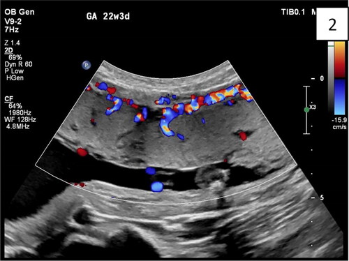
Figure 3. Sagittal transabdominal plan of abdominal invasive anterior placenta with interruption together with a uterus: serosa bladder face with irregular shape with different size, grade 3 lacunae, according to Finberg classification. The abnormal lacunae are mostly at the lower uterine segment under the hyperechoic line between the uterus and bladder. There are four typical markers: loss of ‘clear zone,’ abnormal placental lacunae, bladder wall interruption, and myometrial thinning.
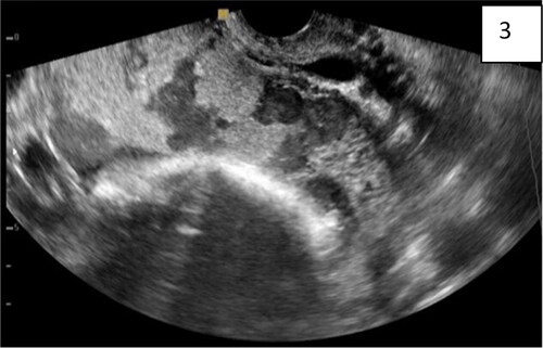
Figure 4. (Same findings as ): transvaginal sonography, abdominal lacunae grade III; specific wall of ‘clear space’ around the cervix (red arrow); subplacental hypervascularity, placenta previa totalis: anterior and posterior.
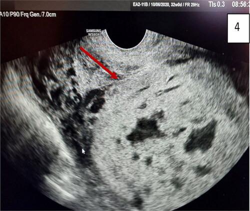
Figure 5. Gray-scale and 3D ultrasound with Power Doppler: multiple images of placenta accreta with bridging vessels on Color Doppler; loss of regularity and interruption of uterine serosa bladder face, demonstrated on 3D. (red arrow).

Figure 6. Ultrasound and pathohistological correlation of total placenta accreta in the lower segment. Lacunae, bridging vessels, and myometrial thinning on ultrasound with loss of anatomical space around the cervix, demonstrated on pathohistological specimen—hysterectomy of the uterus with avoiding of placental space (inset). The placenta is left in situ without separation.
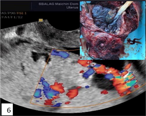
Figure 7. Transvaginal ultrasound of total placenta previa (anterior and posterior) with many ultrasound markers, including the new marker intracervical lakes on Gray-scale; on Color Doppler of the lower uterine segment: bridging vessels, loss of clear space, lacunae with turbulent blood flow inside, feeder vessels and analyzing phenomenon, intraplacental hypervascularity, and intravascular lakes (red arrows).
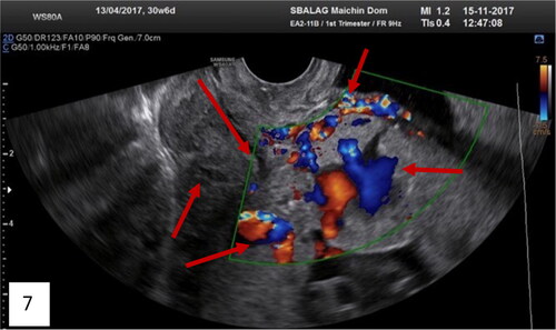
Figure 8. Distraction, placental bulge, and neovascularization of lower uterine segment anteriorly.
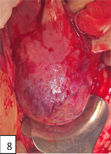
Figure 9. Distraction and neovascularization of lower uterine segment posteriorly.
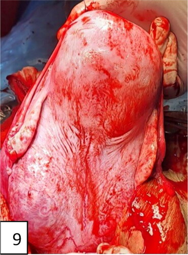
Figure 10. Uterine incision, distraction, extension, and bulging of the lower uterine segment and neovascularization (intraoperative signs of placenta accreta: snowmen sign, distraction, lividity, fading, and ‘jellyfish’—name suggested by M. Tsankova).
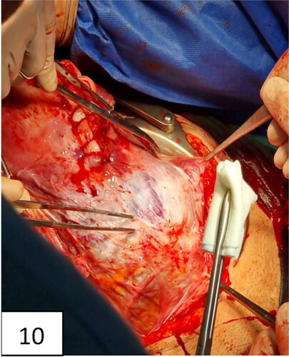
Figure 11. Hysterectomy with signs and markers of , bulging in the lower uterine segment, including the cervix, specific for placenta accreta/percreta.
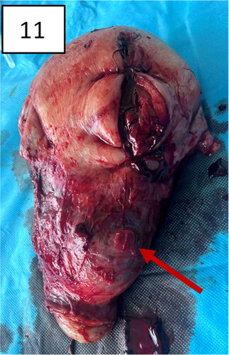
Figure 12. The uterus after hysterectomy with the placenta in situ (pathohistological specimen): thinning of the lower uterine segment and placental tissue, invading the myometrium.
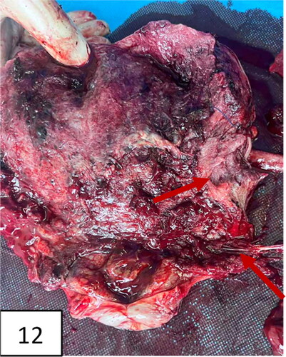
Figure 13. A uterus with invasion of placenta to the uterus cicatrix. Thinning of the cicatrix with loss of myometrium in the zone (arrows). Placenta previa, more anterior and less posterior; vasa previa.
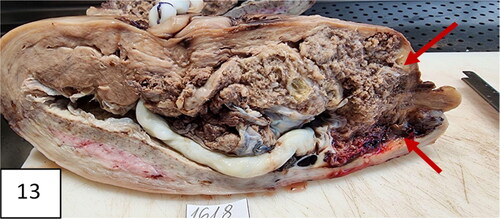
Figure 14. Another slide of uterine specimen: thinning of the cicatrix with loss of myometrium in the zone (arrows). Placenta previa, more anterior and less posterior; vasa previa.
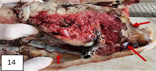
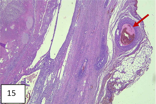
Figure 15. Histological image: chorionic villus with trophoblast invading myometrial muscle (direct contact). Hematoxylin and eosin staining; 200×.
Data availability
All anonymized data that support the findings from this study are available from the corresponding author [N.G.] upon reasonable request.