Figures & data
Figure 1. Surfaces and transverse sections of soybean hypocotyls after 6 d of flooding. The surfaces were photographed under a stereoscopic microscope (A–E and P–S). The sections were excised from hypocotyls at 1.5 cm above the sand surface, and then photographed under a stereoscopic microscope (F–J and T–W) and a light microscope (K–O and X–α). Five plants were used in each treatment. The photographs are surfaces and typical sections from soybean plants grown without flooding (control; A, F, and K) or under flooding with water (B, G, and L), TIBA (C, H, and M), NPA (D, I, and N), HFCA (E, J, and O), 1-NOA (P, T, and X), CHPAA (Q, U, and Y), MH (R, V, and Z) or PCIB (S, W, and α). ae: aerenchyma, co: cortex, pf: phloem fiber, ph: phellogen, xy: xylem. Scale bar = 2 mm (A–E and P–S), 1 mm (F–J and T–W), or 0.2 mm (K–O and X–α).

Table 1. Areas of stele, secondary aerenchyma, cortex, and total, as well as the Ae/St ratio in cross sections of soybean hypocotyls.
Figure 2. Primary leaf epinasty induced by TIBA, HFCA, and NPA under flooding. The plants were photographed after 3 d of treatment. Three to five plants were used in each treatment.
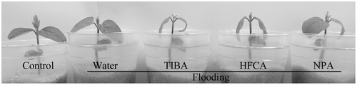
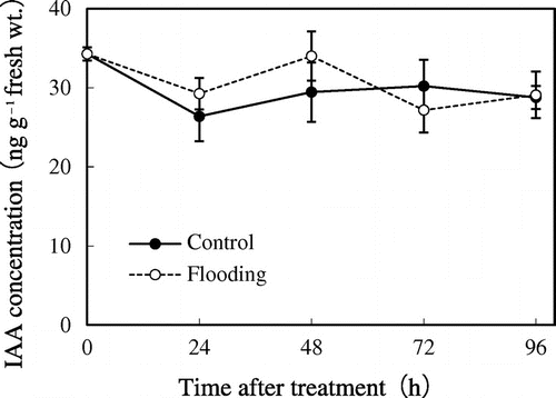
Figure 3. Endogenous IAA concentrations in soybean hypocotyls after flooding. The hypocotyl sections were excised at 0, 24, 48, 72, and 96 h after flooding. Values are shown as means (±SE, n = 4–10). There is no statistical difference in IAA concentration between control and flooded plants at any sampling time as determined by Student’s t-test (p < 0.05).