817
Views
18
CrossRef citations to date
0
Altmetric
Research Article

Homology Model of Human Retinoic Acid Metabolising Enzyme Cytochrome P450 26A1 (CYP26A1): Active Site Architecture and Ligand Binding

, , , , &
Pages 361-369 | Received 01 Mar 2006, Accepted 31 Mar 2006, Published online: 04 Oct 2008

Figures & data

Figure 1 Alcohol dehydrogenases (ADH) and short-chain dehydrogenase/reductase catalyse the oxidation of retinol to retinaldehyde, which is subsequently oxidised by aldehyde dehydrogenases (ALDH) to retinoic acid. Retinoic acid, all-trans (atRA) and 9-cis (9cRA) isomers are further metabolised by cytochrome P450 enzymes (including CYP26A1, B1 and C1) to inactive polar metabolites resulting in retinoid excretion.
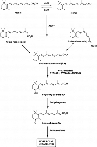
Figure 1 Alcohol dehydrogenases (ADH) and short-chain dehydrogenase/reductase catalyse the oxidation of retinol to retinaldehyde, which is subsequently oxidised by aldehyde dehydrogenases (ALDH) to retinoic acid. Retinoic acid, all-trans (atRA) and 9-cis (9cRA) isomers are further metabolised by cytochrome P450 enzymes (including CYP26A1, B1 and C1) to inactive polar metabolites resulting in retinoid excretion.

Table I.  P450 sequences identified by PSI-BLAST search using CYP26A1 as the query sequence.

Figure 2 CYP26A1 sequence and ClustalW (1.82) alignment with CYP2C9, CYP2C8 and CYP3A4. The colour coding of amino acid type: red, small + hydrophobic (AVFPMILWY); blue, acid (DE); magenta, basic (RHK); green, hydroxyl + amine + basic (STYHCNGQ). Substrate recognition sites (SRS) and conserved P450 domains (ETLR, PERF and Haem binding domain) are indicated. “*” means that the residues are identical, “:” means that conserved substitutions have been observed, “.” means that semi-conserved substitutions are observed.
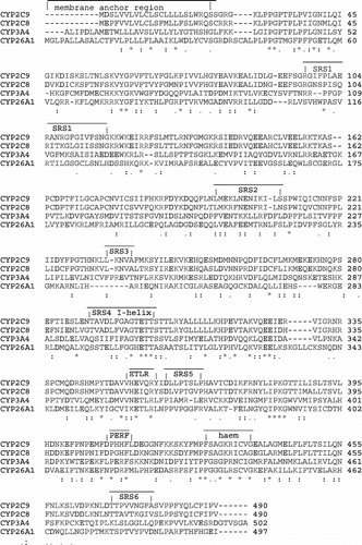
Figure 2 CYP26A1 sequence and ClustalW (1.82) alignment with CYP2C9, CYP2C8 and CYP3A4. The colour coding of amino acid type: red, small + hydrophobic (AVFPMILWY); blue, acid (DE); magenta, basic (RHK); green, hydroxyl + amine + basic (STYHCNGQ). Substrate recognition sites (SRS) and conserved P450 domains (ETLR, PERF and Haem binding domain) are indicated. “*” means that the residues are identical, “:” means that conserved substitutions have been observed, “.” means that semi-conserved substitutions are observed.

Table II.  Validation results for the lowest energy CYP26A1 models (produced from the three different templates) and the 3D structural templates.

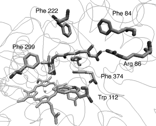
Figure 3 CYP26A1 model active site (a) before and (b) after active site optimisation with the (S)-R115866 bound inhibitor.
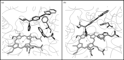
Figure 3 CYP26A1 model active site (a) before and (b) after active site optimisation with the (S)-R115866 bound inhibitor.

Figure 4 Interactions between (S)-R115866 and the CYP26A1 active site.

Figure 4 Interactions between (S)-R115866 and the CYP26A1 active site.

Figure 5 atRA docked in CYP26A1 active site.

Figure 5 atRA docked in CYP26A1 active site.

Table III.  3. Comparison of CYP26A1 model and CYP3A4 template secondary structure.
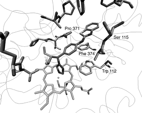
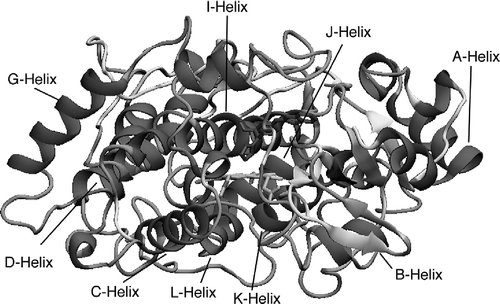
Figure 6 3D Structure of the CYP26A1 model. α-Helices are in red, β-sheets are in yellow, loops are cyan and coils are grey.

Figure 6 3D Structure of the CYP26A1 model. α-Helices are in red, β-sheets are in yellow, loops are cyan and coils are grey.

Reprints and Corporate Permissions

Please note: Selecting permissions does not provide access to the full text of the article, please see our help page How do I view content?

To request a reprint or corporate permissions for this article, please click on the relevant link below:

Academic Permissions

Please note: Selecting permissions does not provide access to the full text of the article, please see our help page How do I view content?

Obtain permissions instantly via Rightslink by clicking on the button below:

If you are unable to obtain permissions via Rightslink, please complete and submit this Permissions form. For more information, please visit our Permissions help page.