Figures & data
Figure 1. Structures of chalcone and compounds I–V.
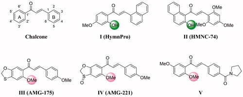
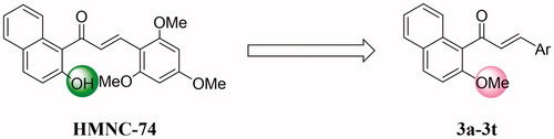
Figure 2. Rationale design of the title compounds of this study.
Scheme 1. Scheme of synthesis of target compounds 3a–3t. Reagents and conditions: (a) Cs2CO3, acetone, r.t. 12 h; (b) 50% KOH (aq), MeOH, 0 °C, 0.5 h to r.t., 24 h.
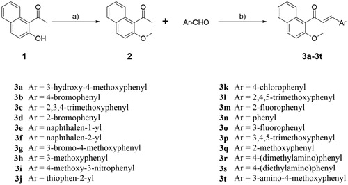
Table 1. Anticancer activity of compounds 3a–3t against MCF-7 cell line.
Table 2. Cytotoxic activity (IC50, µM) of selected compound 3a and cisplatin against human embryonic kidney (HEK293) cell line.
Figure 3. Tubulin polymerisation inhibitory activity of compound 3a (3.0 μM, 6.0 μM, 12.5 μM, and 25.0 μM) and colchicine (12.5 μM).
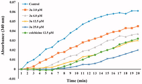
Figure 4. Cell cycle analysis and cell apoptosis analysis for MCF-7 cells. (A,B) Induction of apoptosis by DMSO (control) and compound 3a (2.0 μM); (C,D) Cell cycle analysis of MCF-7 cells after treated with DMSO (control) or compound 3a (2.0 μM) for 24 h.
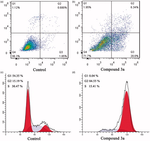
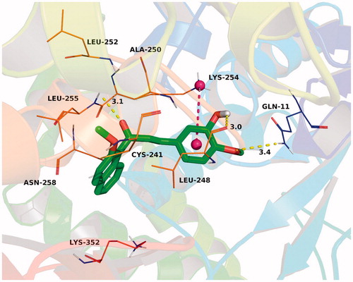
Figure 5. The binding mode of compound 3a (green) with colchicine binding site (magenta) of tubulin (PDB code 1SA0). Hydrogen bonding was depicted as yellow dotted lines.