
Figures & data
Table 1. GenBank numbers from both Trinidadian Chironius taxa. Bolded sequence numbers were generated in this study.
Fig. 1. Phylogenetic relationships of Chironius taxa estimated from a Bayesian 50% majority-rule consensus phylogram using a multilocus dataset (i.e., 12S, 16S, cyt b, ND4, cmos, NT3, Rag-1, Rag-2; total of 5229 bp) with posterior probabilities (≥95) represented at the node (red circles) and additional support values (SH-aLRT > 80%/UFboot <95%) from maximum likelihood (ML) analyses of the partitioned dataset obtained from IQ-TREE. Inserted photographs are top right Chironius nigelnoriegai sp. nov. from Trinidad’s Northern Basin; bottom left is a C. cochranae from the Arima Valley, Trinidad. Photographs by Saifudeen Muhammad and JCM, respectively.
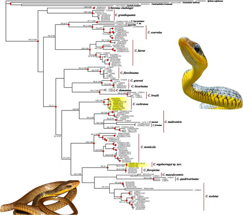
Fig. 2. Divergence time estimates from representative Chironius samples. Inference was based on the combined mtDNA and nDNA data (5229 bp). Red dots indicate mean divergence data. Plio. = Pliocene; Plei. = Pleistocene. Insert is Chironius cochranae from southern Trinidad north of Moruga, photograph by Rainer N. Deo.
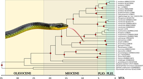
Fig. 3. Holotype of Chironius carinatus Linne 1758, NRM 33. Photograph taken from the Museum Adolphi Friderici collection of the Swedish Museum of Natural History.
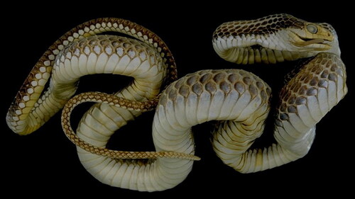
Fig. 4. Chironius cochranae (top) an adult from Petite Tacarib, Trinidad (photograph by Rainer N. Deo); (middle) defensive posture from Mt. Hololo, Trinidad (photograph by Adam Fifi); (bottom) an adult is sleeping high in a tree (photograph by Rainer N. Deo).
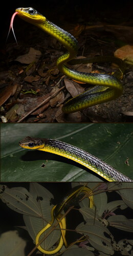
Fig. 5. Comparison of hatchlings. A. Chironius cochranae from Morne Bleu Ridge, Trinidad. B. Chironius nigelnoriegai from the Arima Valley. Note the difference in the size of the eyes. Photographs by JCM.
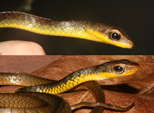
Fig. 6. Illustration of Herpetodryas carinatus reproduced from Jan and Sordelli (Citation1869).
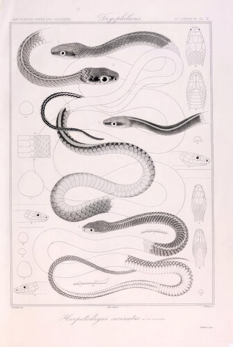
Fig. 7. From top to bottom: Dorsal whole specimen, top of head, right profile, bottom of head, and ventral whole specimen views of the holotype of Chironius nigelnoriegai (CAS 231765). Scale bars equal 1 cm.
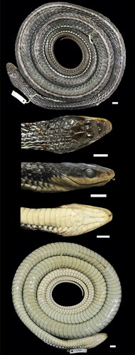
Fig. 8. Chironius nigelnoriegai adult in-life from Manzanilla, Trinidad. Photographs by Adam Fifi.
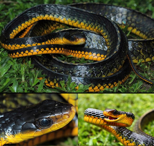
Fig. 9. Head comparison. A–C Chironius cochranae CAS 231763. D–F Chironius nigelnoriegai CAS 231764. Photographs by JCM.
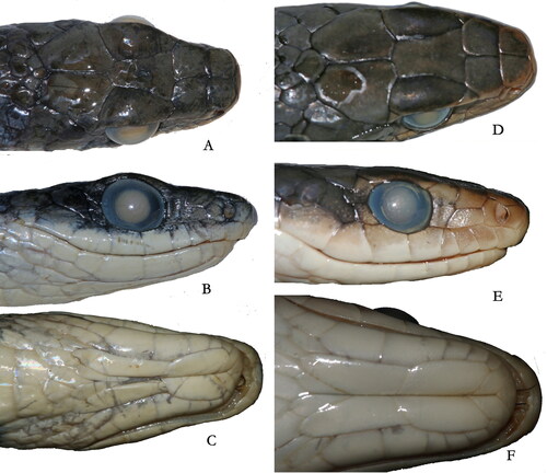
Table 2. Morphological comparison of Chironius nigelnoriegai sp. nov. and C. cochranae. Values for C. cochranae derived from Dixon et al. (Citation1993).