
Figures & data
Figure 1. Cross section of a brain showing grey and white matter.
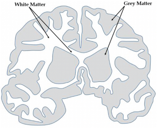
Figure 2. (a) CAD-based mold design for test coupons and (b) 3D printed mold with test coupons.
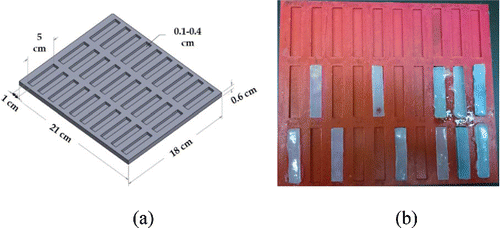
Figure 3. Brain tissue surrogate samples marked for mechanical testing.
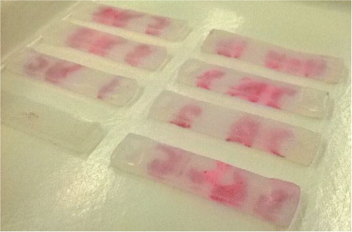
Figure 4. Brain tissue surrogates tested in tension.
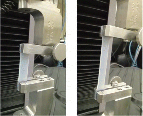
Figure 5. Brain tissue simulant tests and comparison with data from the literature Citation[41].
![Figure 5. Brain tissue simulant tests and comparison with data from the literature Citation[41].](/cms/asset/2105a51e-617d-45d7-aed6-34648b5f4637/umcm_a_1143749_f0005_b.gif)
Table 1. Silicone composition ranges for grey and white matter tissues at low and high strain rates.
Table 2. Control test specimen compositions for mimicking mean brain tissue biomechanical properties and for repeatibility tests.
Figure 6. Brain tissue composition (control specimen 1) mimicking mean white matter mechanical behavior Citation[41] at low strain rate testing, tested for repeatibility.
![Figure 6. Brain tissue composition (control specimen 1) mimicking mean white matter mechanical behavior Citation[41] at low strain rate testing, tested for repeatibility.](/cms/asset/51689ec3-e1f6-4c14-b8b5-26180d33258a/umcm_a_1143749_f0006_b.gif)
Figure 7. Brain tissue composition (control specimen 2) mimicking mean grey matter mechanical behavior Citation[41] at low strain rate testing, tested for repeatibility.
![Figure 7. Brain tissue composition (control specimen 2) mimicking mean grey matter mechanical behavior Citation[41] at low strain rate testing, tested for repeatibility.](/cms/asset/dfed93e3-88cb-4c21-915b-3d2816e9588e/umcm_a_1143749_f0007_b.gif)
Figure 8. Brain tissue composition (control specimen 3) simulating mean grey and white matter mechanical properties Citation[41] at high strain rate testing (30 mm/s), tested for repeatibility.
![Figure 8. Brain tissue composition (control specimen 3) simulating mean grey and white matter mechanical properties Citation[41] at high strain rate testing (30 mm/s), tested for repeatibility.](/cms/asset/288aabec-799a-48d9-a170-cb1727bacc92/umcm_a_1143749_f0008_b.gif)
Figure 9. Average polynomial trendline fits of the silicone specimens tested at high and low strain rates (based on ) for hyperelastic curve fitting.
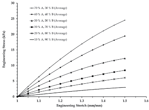
Table 3. Hyperelastic curve fit parameters for the average stress vs. stretch plots of silicone specimens (brain tissue simulants) corresponding to .
Table 4. Accuracy of hyperelastic curve fitting estimated using an average R2 corellation value calculation.