
Figures & data
Figure 1. (a) 5-day incubator control. Note the well-defined pseudostratified ciliated epithelium with goblet cells and underlying basal cells. (b). 5- day mock treatment (air only), 20x. There are 1–2 layers of basal cells with basophilic nuclei, small nucleoli, and amphophilic cytoplasm (red arrow). Up to ∼6 layers of pseudostratified ciliated epithelial cells (asterisk) with darker nuclei and indistinct cytoplasmic borders. There are occasional mucin-producing cells with light blue stippled material (back arrow).
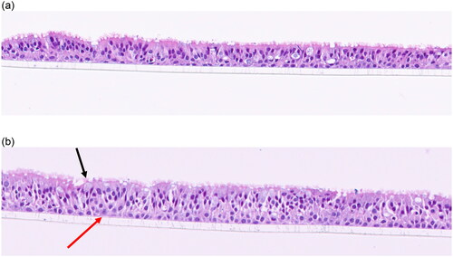
Figure 2. (a). 15-day incubator control. Well-defined pseudostratified ciliated epithelium with goblet cells and underlying basal cells. (b) 15-day mock treatment (air only) control. The respiratory epithelium in the upper panel is thinner (one layer in this image) and the nuclei are darker and more flattened indicative of squamous metaplasia. The dark eosinophilic surface is indicative of keratin production. Basal cells are present adjacent to the scaffold and appear reactive. The Middle panel has more layers of squamous epithelial cells with keratin production and scattered ciliated cells. The lower panel (20+ magnification) illustrates reactive basal cells (arrows) that are larger with paler chromatin and prominent nucleoli.
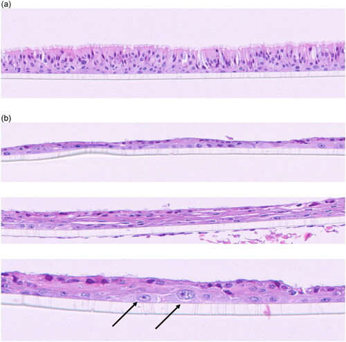
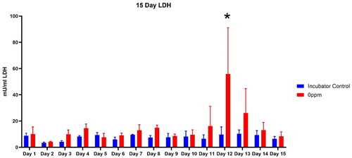
Figure 3. Lactate dehydrogenase release into media from incubator controls and mock-treament air only (0 ppm) VITROCELL® exposure chamber controls.
*LDH in media on Day 12 compared to incubator controls was p < 0.01.
Data availability statement
Data available effective July 15, 2024.