
Figures & data
Figure 1. Tanshinone I inhibits proliferation of Hep3B cells by destabilizing genomes.
(a) The chemical structure of Tanshinone I. (b,c) Representative images (B) and statistical analysis (C) of clonogenic assay which was used to examine the growth of Hep3B cells treated with Tanshinone I at different concentrations. 300 cells per well were seeded in a 6-well plate and Tanshinone I was added according to the following concentrations: 0, 250 nM, 500 nM, 750 nM, 1 μM, 2 μM. ****: P < .0001. (d) Cells treated with Tanshinone I at different concentrations were analyzed for the calculation of the growth rate at different time points using MTT assay. **: P < .0001. ****: P < .0001. (e,f) Representative pictures (E) and statistical analysis (F) of alkaline comet assay. 2 × 10⁴ cells were inoculated into 6-well culture plates, treated with Tanshinone I for 72 h, before being lysed for the analysis of the genomic stability using the comet assay. At least 50 cells per group were quantified using the software of Cometscore. The results were presented as mean ± SEM and Mann-Whitney U test was employed as the method of statistical analysis. ****: P < .0001.
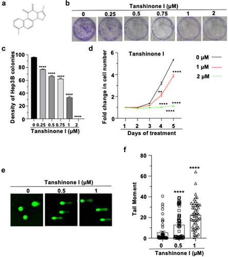
Figure 2. Tanshinone I suppresses the recruitment of γH2AX and inhibits HR and NHEJ repair in Hep3B cells.
(a,b) Representative images of γH2AX foci post IR at different time points (A), and statistical quantifications of the foci number at different points post IR (B). (c) Western blot analysis of γH2AX level in Hep3B cells treated with Tanshinone I at different concentrations. (d,e) The schematic map of the NHEJ (D) and HR (E) reporters for DSB repair analysis. (f,g) Analysis of NHEJ (F) and HR (G) efficiency of cells treated with indicated doses of Tanshinone I. HR and NHEJ reporters were degested with I-SceI in vitro, and transfected, along with DsRed plasmids for normalizing transfection efficiency, into Tanshinone I pre-treated Hep3B cells. On day 3 post transfection, cells were harvested for FACS analysis. *, P < .05, ***: P < .001, ****: P < .0001.
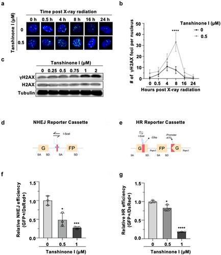
Figure 3. Tanshinone I suppresses the mRNA level of 53BP1 and inhibits the recruitment of RPA2 to DNA damage sites.
(a) Western blot analysis of the expression of NHEJ pathway associated proteins. (b) Real-Time PCR was used to measure the mRNA level of 53BP1. (c-f) Representative images of the foci of 53BP1(C) and RPA2 (E) and statistical quantifications of foci numbers of 53BP1(D) and RPA2 (F) at different time points post X-ray irradiation in Hep3B cells. Hep3B cells were pretreated with Tanshinone I for 72 hours, then irradiated with X-ray at a dosage of 2 Gy. Cells were harvested at indicated time points for immunostaining experiments. Images were taken by a laser confocal microscope (TCS SP8; Leica,wetzla,Germany). At least 50 cells for each time point were quantified.
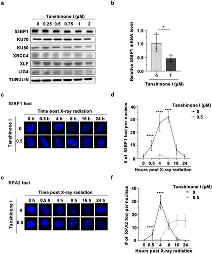
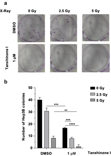
Figure 4. Tanshinone I sensitizes HCC cells to ionizing radiation.
(a) The representative pictures of clonogenic assay showing the survival of hepatoma cells after X-ray irradiation together with Tanshinone I treatment. (b) The statistical analysis of (A). Hep3B cells were pretreated with 0 or 1 μM Tanshinone I for 24 h before being irradiated with X-ray at 0, 2.5, and 5 Gy, followed by the continued Tanshinone I incubation for ~ 2 weeks. The error bars represent S.D.
Supplemental material
Supplemental Material
Download MS Word (151.3 KB)Data availability statement
All data generated during this study are included in the main text and supplementary information files.