
Figures & data
Figure 1. CCT6A expression. CCT6A mRNA expression (a) and protein expression (b, c) were increased in OSCC cells compared to control cells.
Figure 2. CCT6A induced OSCC malignant behaviors. CCT6A mRNA expression (a) and protein expression (b, c) after transfection into SCC-15 cells. Effect of CCT6A on cell proliferation (d), cell apoptosis rate (e, g) and invasive cell count (f, h) in SCC-15 cells. CCT6A mRNA expression (i) and protein expression (j, k) after transfection into HSC-3 cells. Effect of CCT6A on cell proliferation (i), cell apoptosis rate (m, o) and invasive cell count (n, p) in HSC-3 cells.
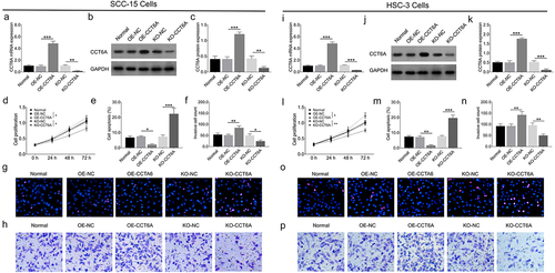
Figure 3. CCT6A promoted OSCC stemness. Effect of CCT6A on sphere formatted number (a, b), CD133 and sox expressions (c, d) in SCC-15 cells. Effect of CCT6A on sphere formatted number (e, f), CD133 and sox expressions (g, h) in HSC-3 cells.
Figure 4. Cross analysis. Cross analysis (a) of accordant DEGs. Further GO enrichment (b) and KEGG enrichment (c) analyses for cross analysis.
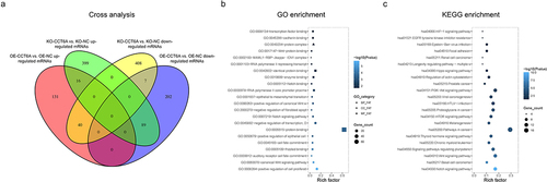
Table 1. Top 50 accordant DEGs.
Table 2. Top 5 pathways.
Figure 5. CCT6A activated Wnt and Notch pathways in OSCC. Effect of CCT6A on Wnt and Notch pathways in SCC-15 cells (a) and in HSC-3 cells (b).
Figure 6. Notch1 and Wnt4 compensated the effect of CCT6A on regulating Notch and Wnt pathways. CCT6A mRNA expression (a) Notch1 mRNA expression (b), Wnt4 mRNA expression (c) and their protein expressions (d, e) in rescue experiments.
Figure 7. Notch1 and Wnt4 compensated the effect of CCT6A knockout on regulating OSCC malignant behaviors. Notch1 and Wnt4 attenuated the effect of CCT6A knockout on regulating cell proliferation (a), cell apoptosis rate (b, d) and invasive cell count (c, e) in SCC-15 cells.
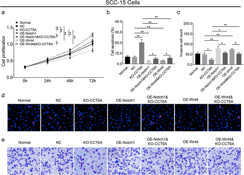
Figure 8. Notch1 and Wnt4 compensated the effect of CCT6A knockout on regulating OSCC stemness. Notch1 and Wnt4 attenuated the effect of CCT6A knockout on regulating sphere formatted number (a, b), CD133 and sox expressions (c, d) in SCC-15 cells.
Supplementary Figure 1.tif
Download TIFF Image (28.9 MB)Supplementary Table 1.docx
Download MS Word (15.2 KB)Supplementary Table 2 clean.docx
Download MS Word (15.7 KB)Data availability statement
The data used to support the findings of this study have been included in this article. The datasets and materials used in this study are available from the corresponding author upon reasonable request.