
Figures & data
Figure 1. Isolation and identification of exosomes from bladder cancer cells.
Exosomes were isolated from SV-HUC-1 (SV-HUC-1-exo), T24 (T24-exo) and UM-UC-3 (UM-UC-3-exo) cells. (a,b) The microscopy and diameter of exosomes were identified by transmission electron microscopy (TEM) and Flow nano analysis. (c) Western blotting detection of exosome markers CD63, CD81, TSG101, and Calnexin in exosomes and cells. Each cell lysate was used as the positive loading control. Three independent experiments were performed for each cell line. Scale bar: 100 nm. TEM: transmission electron microscopy.
Figure 2. Effect of bladder cancer cells-derived exosomes on angiogenesis.
HUVECs were stimulated with SV-HUC-1-exo, T24-exo, and UM-UC-3-exo for 48 h. PBS treatment was performed as a control group. (a) The exosomes were stained by PKH67 and incubated with HUVECs to evaluate the internalization of exosomes. (b) CCK-8 assay was conducted to measure the viability of HUVECs. (c) Representative micrographs of cell migration are determined by transwell assay. Migrated cells were quantified. (d) Representative images of the tube formation assay and quantification of the number of tubes. (e) VEGF level in the supernatants of HUVECs was assessed by ELISA. Three independent experiments were performed for each cell line. *p < 0.05, **p < 0.01, ***p < 0.001. HUVECs: human umbilical vein endothelial cells; DAPI: 4’,6-diamidino-2-phenylindole.
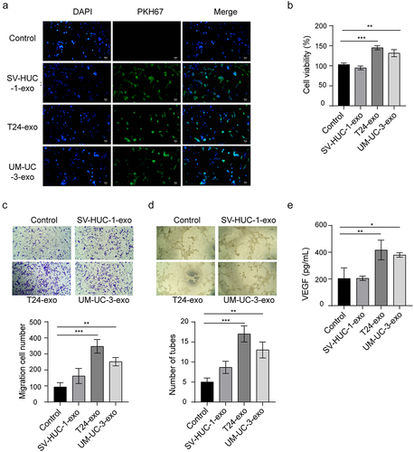
Figure 3. The role of bladder cancer cells-derived exosomal miR-1247-3p in angiogenesis.
HUVECs were treated with SV-HUC-1-exo, T24-exo, and UM-UC-3-exo for 48 h. The control group was treated with PBS. (a) miR-1247-3p expression in exosomes and HUVECs were detected by qRT-PCR. (b) miR-1247-3p inhibitor or inhibitor NC was transfected into T24 and UM-UC-3 cells followed by isolation of the exosomes (miR-1247-3p inhibitor-exo and inhibitor NC-exo) from the supernatants. HUVECs were stimulated with miR-1247-3p inhibitor-exo and inhibitor NC-exo for 48 h. PBS treatment was used as a negative control. Relative miR-1247-3p expression in HUVECs of each group was detected. CCK-8 assay (c), transwell assay (d) and tube formation assay (e) were carried out to measure cell ability, migration, and tube formation ability of HUVECs. (f) ELISA was performed to examine the VEGF level in the supernatants of HUVECs. Three independent experiments were performed for each cell line. *p < 0.05, **p < 0.01,***p < 0.001. HUVECs: human umbilical vein endothelial cells.
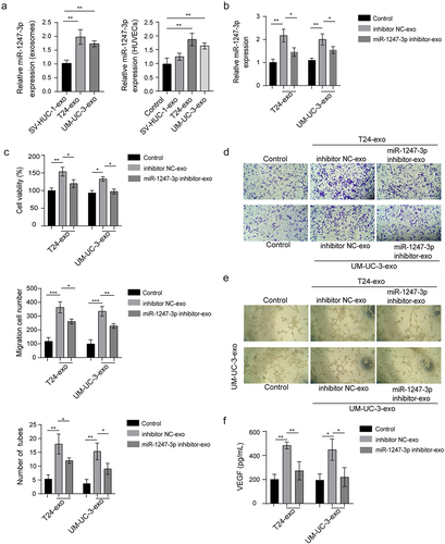
Figure 4. miR-1247-3p targets FOXO1 directly in bladder cancer cells.
(a) Schematic diagram of FOXO1 3ʹ-UTR and the predicted target site of miR-1247-3p. Reporter plasmids containing FOXO1-wt or FOXO1-mut at miR-1247-3p sequence were co-transfected into T24 or UM-UC-3 cells with miR-1247-3p inhibitor or inhibitor NC. The luciferase activity of FOXO1 3ʹ-UTR was examined by dual-luciferase reporter analysis. (b) The target interaction of miR-1247-3p and FOXO1 was confirmed by RNA pull‐down assay. Endogenous FOXO1 enrichment in biotinylated immunoprecipitations was measured by qRT-PCR detection. (c, d) miR-1247-3p inhibitor or inhibitor NC was transfected into T24 or UM-UC-3 cells. FOXO1 mRNA and protein levels were detected by qRT-PCR and western blot. (e, f) Exosomes derived from supernatants of miR-1247-3p inhibitor-transfected cells were co-cultured with HUVECs. FOXO1 mRNA and protein levels in HUVECs were detected. Three independent experiments were performed for each cell line. *p < 0.05, **p < 0.01, ***p < 0.001. wt: wild type; mut: mutation; HUVECs: human umbilical vein endothelial cells.

Figure 5. miR-1247-3p regulates angiogenesis through targeting FOXO1.
(a,b) si-FOXO1 or si-NC was transfected into HUVECs. The mRNA and protein levels of FOXO1 were measured by qRT-PCR and western blot. (c) miR-1247-3p inhibitor and si-FOXO1 were transfected or co-transfected into HUVECs. CCK-8 assay, transwell assay (d) and tube formation assay (e) were conducted to evaluate cell ability, migration, and tube formation ability. (f) ELISA was used to measure the VEGF level in the supernatants. Three independent experiments were performed for each cell line. *p < 0.05, **p < 0.01. HUVECs: human umbilical vein endothelial cells.
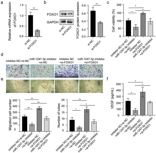
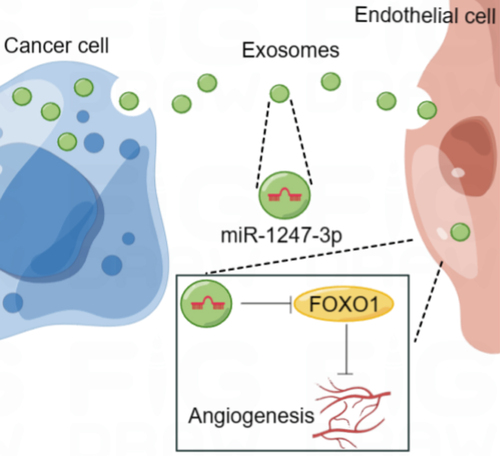
Figure 6. Schematic illustration of the functional role and potential mechanism of exosomal miR-1247-3p in angiogenesis of bladder cancer (by figdraw).
Supplemental material
Supplemental Material
Download MS Word (39.7 KB)Data availability statement
The datasets used or analyzed during the current study are available from the corresponding author on reasonable request.