
Figures & data
Table 1. shRnas targeting sequences.
Figure 1. Knockdown of DIRAS1 significantly promotes proliferation, growth and motility of C33A and SiHa cells.
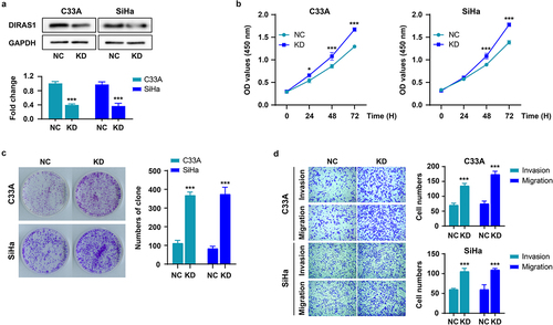
Figure 2. DIRAS1 overexpression significantly inhibits the proliferation, growth and motility of C33A and SiHa cells.
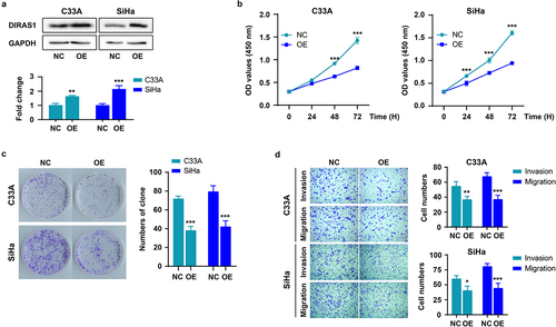
Figure 3. Altered expression pattern of DIRAS1 in cervical cancer tissues.
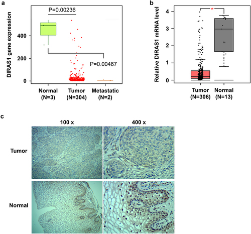
Table 2. DIRAS1 expression in cervical cancer tissues compared with para-carcinoma tissues.
Table 3. DIRAS1 expression associated with the clinicopathological parameters in CCA.
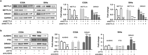
Figure 4. The mechanisms of epigenetic regulation to regulate DIRAS1.
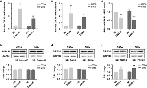
Figure 5. M6A modification promotes DIRAS1 translation.
Data availability statement
The datasets generated during and/or analyzed during the current study are not publicly available, but are available from the corresponding author on reasonable request.