
Figures & data
Figure 1. Higher expression of SUMOylation-related molecules are associated with increased genomic instability and double minute (DM) counts.
(a) SUMOylation-related molecules, such as UBC9 and small ubiquitin‑related modifier (SUMO) 1–3, were more highly expressed in colorectal cancer samples, from the cBioPortal database, with greater genomic instability, namely microsatellite (MSI, n = 60) and chromosome instability (CIN, n = 226), compared to genome stability group (GS, n = 49). (b) UBC9 and SUMO1-3 expression levels were higher among breast cancer samples classified as “DMs” (n = 97), compared to “no DMs” (n = 190), obtained from The Cancer Genome Atlas (TCGA). (c) SUMOylation-related molecule UBC9, and SUMO2 expression levels were significantly higher among “DMs++” group (n = 13), compared to “no DMs” (n = 50) and “DMs+” (n = 29), based on karyotypes from the Cancer Cell Line Encyclopedia (CCLE) database. (d) SUMO1-2, SAE2, and UBC9 were more highly expressed among both DM cell lines, from ovarian cancer-derived UACC-1598DM/UACC-1598HSR and colorectal cancer-derived Colo320DM/Colo320HSR, under RT-qPCR. (e) Western blot showing increased protein expression of UBC9 among both DM cell lines, compared to non-DM ones, but not for SAE2. Student’s t-test was used for comparisons between two groups and one-way analysis of variance (ANOVA) with Tukey’s post hoc test for three or more groups. *P < .05, **P < .01, and ***P < .001.
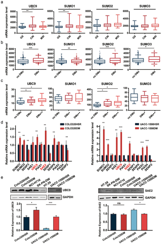
Figure 2. UBC9 knockdown is associated with lowered DMs and DM-carried gene expression.
(a) Western blot showing successful UBC9 knockdown post-short hairpin RNA (shRNA) transfection among UACC-1598DM cells. (b) Karyotypes showing reduced DM counts after UBC9 knockdown in UACC-1598DM. (c) Copy numbers for DM-carried oncogenes MYCN, EIF5A2, and MCL-1 in UACC-1598DM significantly decreased upon UBC9 knockdown. (d) Western blot showing that protein expression levels for EIF5A2 and MYCN significantly decreased upon UBC9 knockdown in UACC-1598DM. (e) Western blot showing successful UBC9 knockdown post-shRNA transfection among Colo320DM cells. (f) Karyotypes showing reduced DM counts after UBC9 knockdown in Colo320DM. (g) Copy numbers for DM-carried oncogenes CDX2, FAM84B, and MYC significantly decreased after UBC9 knockdown in Colo320DM. (g) Western blot showing that protein expression levels for MYC and CDX2 significantly decreased upon UBC9 knockdown in Colo320DM. One-way ANOVA with Tukey’s post hoc test was used for comparisons among the groups. *P < .05, **P < .01, and ***P < .001.
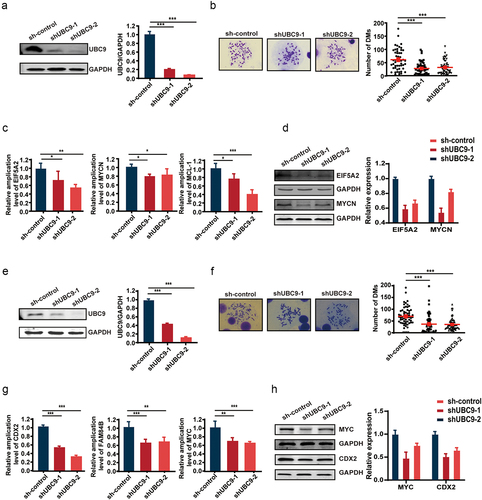
Figure 3. UBC9 inhibition by the 2-D08 inhibitor yields similar results as UBC9 knockdown, due to reduced SUMOylation.
(a) SUMO1/2 expression was significantly lower among UACC-1598DM cells exposed to 2-D08 (30 µM) for 7 d, compared to DMSO control. (b) Karyotypes showing reduced DM counts among UACC-1598DM after 2-D08 treatment versus DMSO control. (c) Copy numbers and (d) mRNA expression levels for DM-carried oncogenes EIF5A2, MCL-1, and MYCN in UACC-1598DM significantly decreased upon UBC9 inhibition. (e) Western blot showing that protein expression levels for EIF5A2 and MYCN significantly decreased upon UBC9 inhibition in UACC-1598DM. (f) Karyotypes showing reduced DM counts among Colo320DM after 2-D08 treatment versus DMSO control. (g) Copy numbers and (h) mRNA expression levels for DM-carried oncogenes FAM84B, MYC, and CDX2 in Colo320DM significantly decreased upon UBC9 inhibition. (i) Western blot showing that protein expression levels for CDX2 and FAM84B significantly decreased upon UBC9 inhibition in Colo320DM. Student’s t-test was used for comparisons between two groups. *P < .05, **P < .01, and ***P < .001.
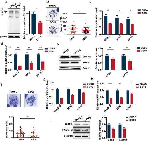
Figure 4. UBC9 knockdown reduces tumor malignancy and growth.
(a) UBC9 knockdown (shUBC9-1 and -2) lowered cell proliferation rates for both UACC-1598DM (left) and Colo320DM cell lines (right), compared to the control group (sh-control). (b) UACC-1598DM cell colonies significantly decreased after UBC9 knockdown. (c) Cell migration for UACC-1598DM (left) and Colo320DM (right) significantly decreased post-UBC9 knockdown. (d) UBC9 was highly expressed in multiple tumors, such as cholangiocarcinoma (CHOL), diffuse large B cell lymphoma (DLBC), glioblastoma multiforme (GBM), pancreatic adenocarcinoma (PAAD), stomach adenocarcinoma (STAD), and thymoma (THYM), under the GEPIA database, compared to normal samples. (e) Higher UBC9 expression was associated with worse survival among ovarian and colorectal cancer patients under Kaplan-Meier Plotter analysis of GSE51373, GSE63885, GSE30161 (ovarian), and GSE41258 (colorectal) datasets. Student’s t-test was used for comparisons between two groups and one-way ANOVA with Tukey’s post hoc test for three or more groups. *P < .05, **P < .01, and ***P < .001.
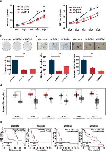
Figure 5. UBC9 inhibition in vivo decreases the rate of tumor growth, via eliminating DMs.
(a) Schematic outlining UACC-1598DM cell injection within 6-week-old SCID female mice to form stable transplanted tumors, as well as subsequent 2-D08 injections (b) Photographs of tumors obtained from sacrificed mice after the end of the experiment, on Day 27. Tumor volumes (c) and weights (d) obtained from both 2-D08-injected mice and DMSO controls. (e) Karyotypes showing reduced DM counts within isolated tumor cells after 2-D08 treatment, compared to DMSO control, among three mouse pairs. (f) Copy numbers and (g) mRNA expression levels for DM-carried oncogenes EIF5A2, MCL-1, and MYCN, among 2-D08-treated mice, were significantly lower than for DMSO control. (H) Western blot showing that protein expression levels for EIF5A2 and MYCN significantly decreased among 2-D08-treated mice, compared to DMSO control. Student’s t-test was used for comparisons between two groups. *P < .05, **P < .01, and ***P < .001.
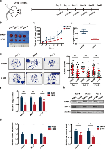
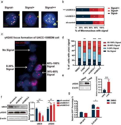
Figure 6. Lowered UBC9 activity is associated with increased micronucleus (MN) expulsion, DNA damage, and decreased DNA damage repair.
(a) Classification of UACC-1598DM cells into three categories, based on the extent of DM signal detected within expelled MN detected by fluorescence in situ hybridization: Signal-, Signal+ and Signal++. (b) UBC9 knockdown (shUBC9-1 and -2) was associated with increased expulsion of DM-containing MNs, compared to control (sh-control). (c) Classification of “No Signal”, “0–30% Signal”, “30–60% Signal”, and “60–100% Signal”, based on the percentage area of γ-H2AX signal, a marker of DNA damage, within the whole nucleus. (d) UBC9 inhibition or knockdown, compared to DMSO or sh-control, had significantly larger areas of “60–100% Signal”, and thus DNA damage, present. Western blot analysis showing that (e) UBC9 inhibition and (f) knockdown increased γ-H2AX expression. (g) UBC9 inhibition significantly decreased non-homologous end-joining (NHEJ) DNA repair, as demonstrated by significantly lower fluorescence intensity in the pimEJ5GFP-transfected cell line, representing NHEJ. By contrast, no significant difference was found for pHPRT-DRGFP-transfected cell line, representing homologous recombination repair. Student’s t-test was used for comparisons between two groups and one-way ANOVA with Tukey’s post hoc test for three or more groups. *P < .05, **P < .01, and ***P < .001.
Supplemental material
Supplemental Material
Download Zip (11.5 MB)Data availability statement
The datasets supporting the conclusion of this article are included within the article.