
Figures & data
Figure 1. LA Inhibited cell proliferation in EC cell lines and tumor growth in a transgenic mouse model.
The HEC-1A and KLE cells were treated with the indicated doses of LA for 72 h. Cell proliferation was detected using an MTT assay. LA inhibited cell growth in a dose-dependent manner in both the cell types (a). The treatment of HEC-1A and KLE cells with LA for 24 h significantly increased the expression of p-ACC (b). The effect of LA on the AMPK and AKT/mTOR pathways in both cell lines was assessed by western blotting. LA treatment significantly increased the expression of p-AMPK and decreased the expression of p-AKT and p-S6 after 24 h of treatment (c). Lkb1fl/flp53fl/fl mice were treated with LA (20 mg/kg, oral, daily) or vehicle for four weeks, and the results showed that LA effectively reduced tumor weight compared with control mice (d). IHC results showed that LA treatment reduced the expression of Ki67 and induced the expression of p-ACC in EC tumor tissues from Lkb1fl/flp53fl/fl mice (e). *p < .05, **p < .01. 200
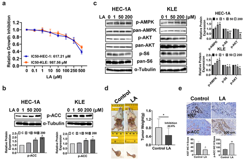
Figure 2. LA Induced cell cycle G1 arrest.
HEC-1A and KLE cells were treated with 1, 50, and 200 μM LA for 36 h, and cell cycle progression was analyzed using a Cellometer. LA induced cell cycle G1 arrest in both cell lines (a). Western blotting was performed to detect the expression of cell cycle-related proteins. LA reduced CDK4 and cyclin D1 expression in both cell lines after 24 h of treatment (b). Cyclin D1 expression was measured by IHC in EC tumors from Lkb1fl/flp53fl/fl mice. Treatment of mice with LA for four weeks significantly decreased the expression of cyclin D1 (c) *p < .05, **p < .01.
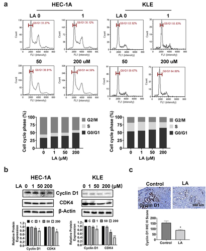
Figure 3. Effect of LA on apoptosis in EC cells and Lkb1fl/flp53fl/fl mice.
Cleaved caspase 3, 8, and 9 activities were determined using ELISA. After treatment with 1, 50, and 200 μM LA for 16 h, the activities of cleaved caspase 3, 8, and 9 were increased in a dose-dependent manner in both HEC-1A and KLE cell lines (a). The expression of Mcl-1 and Bcl-xL decreased in both cell lines after treatment with LA for 24 h (b). LA inhibited the expression of Bcl-xL in EC tumor tissues, as detected by IHC (c).*p < .05, **p < .01.
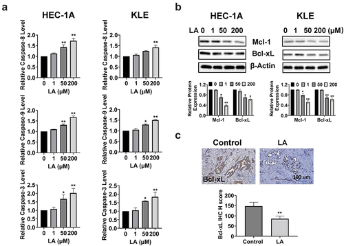
Figure 4. Effect of LA on cellular stress in EC cells and Lkb1fl/flp53fl/fl mice.
HEC-1A and KLE cells were treated with 1, 50, and 200 µM LA for 14 h. LA significantly increased the ROS levels in both cell lines (a). The TMRE assay showed that 200 µM LA effectively decreased mitochondria membrane potential in HEC-1A and KLE cells (b). The TEAC assay demonstrated that 50 and 200 µM LA significant reduced total antioxidant capacity in both cells after 24 hours of treatment (c). Western blotting results revealed that LA increased the expression of BiP and PERK proteins after treatment with LA for 14 h (d). IHC staining showed an increase in the expression of BiP in LA-treated Lkb1fl/flp53fl/fl mice compared with that in control mice (e). *p < .05, **p < .01.
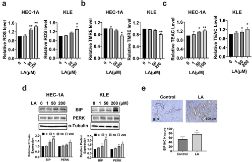
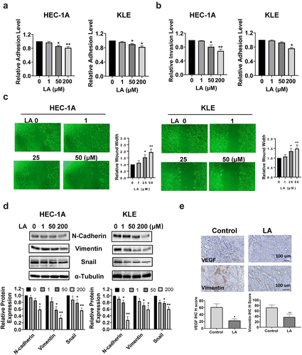
Figure 5. LA Inhibited adhesion and invasion.
Adhesive and invasive abilities were detected by laminin-1 and transwell assays in EC cell lines, respectively. LA inhibits cell adhesion and invasion in HEC-1A and KLE cells (a,b). The wound healing assay showed that cell migration was inhibited by LA after 48 h of treatment in both cell lines (c). Western blotting showed a descending trend in the expression of EMT-related proteins, including N-Cadherin, vimentin, and snail (d). IHC results indicated that treatment with LA for four weeks in Lkb1fl/flp53fl/fl mice inhibited the expression of VEFG and vimentin in EC tumor tissues compared with that in control mice (e).*p < .05, **p < .01.
Data availability statement
All data generated or analyzed during this study are included in this article. The datasets used and/or analyzed during the current study are available from the corresponding authors upon reasonable request.