Figures & data
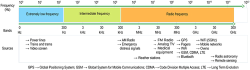
Table 1. EMF characteristics in the extremely low-frequency range (0–300 Hz) utilized to expose assorted cell lines, along with subsequent biological effects.
Table 2. EMF characteristics in the intermediate frequency range (300 Hz-300 kHz) utilized to expose assorted cell lines, along with subsequent biological effects.
Table 3. EMF characteristics in the radio frequency range (300 kHz-300 GHz) utilized to expose assorted cell lines, along with subsequent biological effects.
Ashdown CP, Johns SC, Aminov E, et al. Pulsed low-frequency magnetic fields induce tumor membrane disruption and altered cell viability. Biophys J. 2020;118(7):1552–1563. Oh IR, Raymundo B, Jung SA, et al. Extremely low-frequency electromagnetic field altered PPARγ and CCL2 levels and suppressed CD44+/CD24- breast cancer cells characteristics. Bull Korean Chem Soc. 2020;41(8):812–823. Vincenzi F, Ravani A, Pasquini S, et al. Pulsed electromagnetic field exposure reduces hypoxia and inflammation damage in neuron-like and microglial cells. J Cell Physiol. 2017;232(5):1200–1208. Buckner CA, Buckner AL, Koren SA, et al. Exposure to a specific time-varying electromagnetic field inhibits cell proliferation via cAMP and ERK signaling in cancer cells. Bioelectromagnetics. 2018;39(3):217–230. Storch K, Dickreuter E, Artati A, et al. Bemer electromagnetic field therapy reduces cancer cell radioresistance by enhanced ros formation and induced DNA damage. PLoS ONE. 2016;11(12):e0167931. Akbarnejad Z, Eskandary H, Vergallo C, et al. Effects of extremely low-frequency pulsed electromagnetic fields (ELF-PEMFs) on glioblastoma cells (U87. Electromagn Biol Med. 2017;36(3):238–247. Liu Y, Liu W, Liu K, et al. Overexpression of miR-26b-5p regulates the cell cycle by targeting CCND2 in GC-2 cells under exposure to extremely low frequency electromagnetic fields. Cell Cycle. 2016;15(3):357–367. Destefanis M, Viano M, Leo C, et al. Extremely low frequency electromagnetic fields affect proliferation and mitochondrial activity of human cancer cell lines. Int J Radiat Biol. 2015;91(12):964–972. Garg AA, Jones TH, Moss SM, et al. Electromagnetic fields alter the motility of metastatic breast cancer cells. Commun Biol. 2019;2(1):1–16. Berkelmann L, Bader A, Meshksar S, et al. Tumour-treating fields (TTFields): investigations on the mechanism of action by electromagnetic exposure of cells in telophase/cytokinesis. Sci Rep. 2019;9(1):1–11. Neuhaus E, Zirjacks L, Ganser K, et al. Alternating electric fields (TTFields) activate Cav1.2 channels in human glioblastoma cells. Cancers (Basel). 2019;11(1):110. Peng WY, Li KJ, Xie YZ, et al. Development of a TEM-cell-integrated CO2 incubator for cell-based transient electromagnetic field bioeffect study. Electromagn Biol Med. 2020;39(4):290–297. Jimenez H, Wang M, Zimmerman JW, et al. Tumour-specific amplitude-modulated radiofrequency electromagnetic fields induce differentiation of hepatocellular carcinoma via targeting Cav3.2 T-type voltage-gated calcium channels and Ca2+ influx. EBioMedicine. 2019;44:209–224. Jooyan N, Goliaei B, Bigdeli B, et al. Direct and indirect effects of exposure to 900 MHz GSM radiofrequency electromagnetic fields on CHO cell line: evidence of bystander effect by non-ionizing radiation. Environ Res. 2019;174:176–187. Choi J, Min K, Jeon S, et al. Continuous exposure to 1.7 GHz LTE electromagnetic fields increases intracellular reactive oxygen species to decrease human cell proliferation and induce senescence. Sci Rep. 2020;10(1):1–15. Sueiro-Benavides RA, Leiro-Vidal JM, Salas-Sánchez AÁ, et al. Radiofrequency at 2.45 ghzGhz increases toxicity, pro-inflammatory and pre-apoptotic activity caused by black carbon in the RAW 264.7 macrophage cell line. Sci Total Environ. 2021;765:142681. Mumtaz S, Bhartiya P, Kaushik N, et al. Pulsed high-power microwaves do not impair the functions of skin normal and cancer cells in vitro: a short-term biological evaluation. J Adv Res. 2020;22:47–55.