Figures & data
Table 1. List of primers used in the study
Figure 1. NRIR is expressed in response to bacterial infection
(a) The expression levels of a few of the annotated non-coding transcripts was analysed by qRT PCR in THP1 macrophages infected with Mtb for 24 hr. The change in expression in infected macrophages relative to the uninfected macrophage levels (log2FC) is depicted as a heat map. Three independent replicate experiments are individually represented as mean expression values from triplicate assays (N = 3). GAPDHwas used as a control for expression analysis.(b–e) Expression of NRIR (b–e) and NEAT1 (b) in macrophages infected with Mtb (b–d) or E. coli/ -Ec or Salmonella – St (e) at different time intervals was analysed by qPCR. Change in expression in infected macrophages (I) relative to uninfected (U) macrophages is represented from triplicate assays of N = 2–3 replicate experiments. U- Uninfected, H- Heat killed Mtb, B- M. bovis BCG.
Figure 2. Identification of the novel transcripts-TILT encompassing CMPK2 and lncRNA – NRIR in Mtb infected macrophages
(a) The genomic locus of CMPK2 and NRIR in human cells is depicted with the previously annotated transcripts. The primers used for PCR analysis of transcripts are depicted by numbers (1–6).(b–e) Analysis of the PCR amplification of TILT with primers spanning the CMPK2-NRIR locus from THP1 cells either uninfected or infected with Mtb for 24 hr. b) TILT amplification from genomic DNA (G) or cDNA (R) of infected sample with primers 1 and 4 (L-DNA ladder). c- PCR amplification of cDNA from uninfected (U) or Mtb infected (I) cells using primers 1 and 4. d- PCR amplification of CMPK2 (3, 4), NRIR (1, 2) and TILT (1,4) by using the 800 bp (open star) and 1 kb (dark star) PCR amplicons as template. e- The sequencing profiles of exons present in the 3 new amplicons.(f) Expression of the novel transcripts in total blood of a healthy individual.
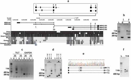
Figure 3. TILT is actively induced in Mtb infected macrophages
(a) Expression kinetics of TILT in THP1 macrophages infected with Mtb at a MOI of 5. At the indicated intervals, RNA prepared from cells was used for analysis by qRT PCR. RNA from THP1 cells Inset- at 6 and 24 hr of infection with Heat killed Mtb (Δ) or BCG.(b) TILT expression in MDMs of two healthy individuals infected with Mtb at a MOI of 5 for 6 and 24 hr (square and circle).(c) TILT expression in blood of healthy individuals (H) and TB patients (P); each symbol represents one individual.(d) TILT expression in THP1 macrophages infected with E. coli (Ec) or Salmonella (St) at a MOI of 5 for the indicated time intervals. Values represented are mean + SEM of triplicate wells of 2–3 independent experiments.The expression of TILT in infected macrophages w.r.t uninfected controls is depicted as rel. expression (a, b, d).
Figure 4. TILT is induced temporally in response to LPS stimulation in THP1 macrophages
(a) TILT expression in macrophages stimulated with different TLR ligands- L- LPS (TLR4) at 10 ng/ml, P- Peptidoglycan (TLR2) at 20 ng/ml, C- Pam3CSK (TLR2/1) at 20 ng/ml, Z-Zymosan (TLR2/6) at 10 µg/ml, IC- poly I:C (TLR3) at 2 µg/ml) at 6 and 24 h post stimulation.(b) Kinetics of TILT and NRIR expression in THP1 macrophages treated with 10 ng/ml of LPS. Relative expression at any given time w.r.t unstimulated cells before stimulation (T0) is depicted as mean ± SEM for triplicate assays of 3 independent experiments.(c) Expression of TILT in LPS stimulated MDMs from PBMCs of two individuals (square and circle).(d) TILT expression in macrophages infected with salmonella (St) (3 hr p.i.), E. coli (Ec) (3 hr p.i.), Mtb (24 hr p.i.) or stimulated with LPS (6 hr p.s.) in the presence of the TLR4 inhibitor CLI095 (3 µM). At the indicated intervals RNA prepared from cells was used for analysis by qRT PCR and normalized to GAPDH.