Figures & data
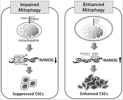
Figure 1. Model of how mitophagy regulates CSCs. When mitophagy is impaired (left panel), PINK1 phosphorylates TP53 at S392, likely on mitochondria, resulting in the activation and nuclear localization of TP53. TP53 then suppresses the expression of NANOG and reduces the self-renewal ability of CSCs. When mitophagy is enhanced (right panel), TP53 associated with PINK1 on mitochondria is degraded with mitochondria in autophagic vacuoles. This leads to the induction of NANOG expression by POU5F1/OCT4 and SOX2, and the enhancement of self-renewal of CSCs.