445
Views
0
CrossRef citations to date
0
Altmetric
Autophagic Punctum

Developmentally regulated selective autophagy determines ER inheritance by gametes

& ORCID Icon
Pages 1732-1733 | Received 20 Jan 2022, Accepted 02 Feb 2022, Published online: 27 Feb 2022

Figures & data

Figure 1. Model of ER inheritance and degradation during meiosis. Top: During meiosis, the cortical ER undergoes a transition from continuous to fragmented, leading to the collapse of most cortical ER. ER fragments that are closely associated with ER-plasma membrane tethers are retained at the cell cortex and excluded from gametes (1). Developmentally timed Atg40 expression mediates the degradation of a subset of collapsed ER by selective autophagy (2), and a subset of collapsed ER is inherited by gametes (3). Bottom: Speculative model for ER quality control during meiosis. We propose that markers of damaged or stressed ER, including lumenal protein aggregates, can be eliminated by selective targeting to cortically retained ER fragments (1) and/or degradation by selective autophagy (2), ensuring that only healthy ER is passed on to gametes (3). Right: Outline of three distinct ER fates during meiosis, highlighting key mediators. Figure adapted from reference 1.
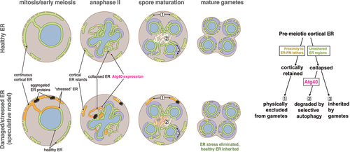
Figure 1. Model of ER inheritance and degradation during meiosis. Top: During meiosis, the cortical ER undergoes a transition from continuous to fragmented, leading to the collapse of most cortical ER. ER fragments that are closely associated with ER-plasma membrane tethers are retained at the cell cortex and excluded from gametes (1). Developmentally timed Atg40 expression mediates the degradation of a subset of collapsed ER by selective autophagy (2), and a subset of collapsed ER is inherited by gametes (3). Bottom: Speculative model for ER quality control during meiosis. We propose that markers of damaged or stressed ER, including lumenal protein aggregates, can be eliminated by selective targeting to cortically retained ER fragments (1) and/or degradation by selective autophagy (2), ensuring that only healthy ER is passed on to gametes (3). Right: Outline of three distinct ER fates during meiosis, highlighting key mediators. Figure adapted from reference 1.

Reprints and Corporate Permissions

Please note: Selecting permissions does not provide access to the full text of the article, please see our help page How do I view content?

To request a reprint or corporate permissions for this article, please click on the relevant link below:

Academic Permissions

Please note: Selecting permissions does not provide access to the full text of the article, please see our help page How do I view content?

Obtain permissions instantly via Rightslink by clicking on the button below:

If you are unable to obtain permissions via Rightslink, please complete and submit this Permissions form. For more information, please visit our Permissions help page.