Figures & data
Figure 5. (E) Capillary-like tube formation was assessed by matrigel angiogenesis assay in HUVECs. HUVECs were cultured either in NG or HG medium in the presence or absence of MET (50 μM) for 72 h, MAN was used as the osmotic control for HG. For pharmacological manipulation of autophagy, HUVECs were treated with RAPA (10 nM) 2 h after MET treatment. Scale bars: 300 μm.
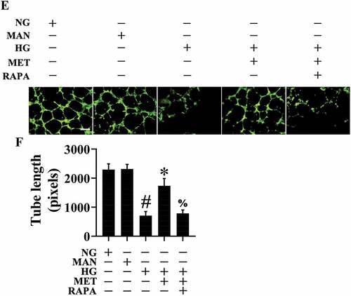
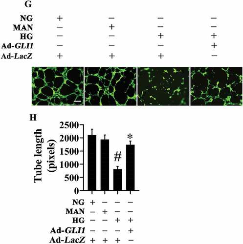
Figure 9. (G) Capillary-like tube formation was assessed by matrigel angiogenesis assay in HUVECs. HUVECs were transduced with adenoviruses harboring GLI1 (Ad-GLI1) and Ad-LacZ (served as a control), respectively. After transduction, HUVECs were cultured either in NG or HG medium for 72 h, MAN was used as the osmotic control for HG. Scale bars: 300 μm.