Figures & data
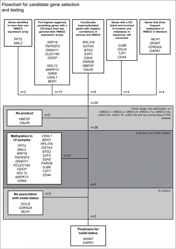
Figure 1. Flowchart for candidate gene selection and testing * TJP1 showed methylation in all samples and was therefore excluded.

Table 1. Selected candidate genes
Table 2. Clinicopathological characteristics
Table 3. Cross table analyses of the 5 genes eligible for testing on the patient series
Table 4. Univariate and multiple logistic regression with pN status
Table 5. (A) Multiple logistic regression of DAPK1 and MGMT for pN status. (B) Crosstable for the DAPK1 and MGMT test combined vs. pN status
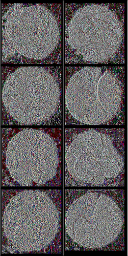
Figure 2. Representative examples of immunohistochemical staining. (A) DAPK1 low expression core, tumor center; (B) DAPK1 high expression, core tumor center; (C) DAPK1 low expression core, tumor front; (D) DAPK1 high expression core, tumor front; (E) MGMT low expression core, tumor center; (F) MGMT high expression core, tumor center; (G) MGMT low expression core, tumor front; (H) MGMT high expression core, tumor front.

Figure 3. Examples of 2 cases that showed MGMT methylation, associated with low expression in the invasive tumor front, but high expression in the tumor center. (A) MGMT methylation controls [pure water, leucocytes, and IV (in vitro SssI methylated leucocytes)] and 2 cases. (B) Low MGMT expression in the tumor invasive front (Case 1). (C) High MGMT expression in the tumor center (Case 1). (D) Low MGMT expression in the tumor invasive front (Case 2). (E) High MGMT expression in the tumor center (Case 2). U: unmethylated; M: methylated; Blanco: pure water control; Leuco: leucocytes; IV: in vitro SssI methylated leucocytes. T: tumor tissue. The border of the tumor area is indicated by a black line.
![Figure 3. Examples of 2 cases that showed MGMT methylation, associated with low expression in the invasive tumor front, but high expression in the tumor center. (A) MGMT methylation controls [pure water, leucocytes, and IV (in vitro SssI methylated leucocytes)] and 2 cases. (B) Low MGMT expression in the tumor invasive front (Case 1). (C) High MGMT expression in the tumor center (Case 1). (D) Low MGMT expression in the tumor invasive front (Case 2). (E) High MGMT expression in the tumor center (Case 2). U: unmethylated; M: methylated; Blanco: pure water control; Leuco: leucocytes; IV: in vitro SssI methylated leucocytes. T: tumor tissue. The border of the tumor area is indicated by a black line.](/cms/asset/7a2a2523-5d08-4944-9e99-b900ef426486/kepi_a_1075689_f0003_oc.gif)
Table 6. Associations between methylation and expression for MGMT and DAPK1
