
Figures & data
Table 1. Autophagic cargos and/or receptors have been identified and characterized for selective autophagy in plants
Figure 1. Cytoplasmic soluble GFP protein is nonselectively engulfed by autophagosomes and delivered into vacuole upon autophagy induction condition
(A) Accumulation of soluble GFP protein in vacuole is dependent on autophagy pathway. Five-day-old Arabidopsis seedlings expressing GFP were treated with ConcA (0.5 μM) for 6 hours and then were subjected to confocal observing. The GFP signal was observed in vacuole upon ConcA treatment, but was not observed in autophagic defective mutant atg7-2. Arrows and arrowheads indicated globular and punctate structures of GFP in vacuole, respectively. Bar = 50 μm. (B) Colocalization between the GFP protein and the autophagic marker mCherry-ATG8f in vacuole. Five-day-old Arabidopsis seedlings co-expressing GFP and mCherry-ATG8f were treated with BTH and ConcA (BTH+ConcA) for 6 hours to induce autophagy, and then were subjected to confocal imaging. Bar = 20 μm. BTH, Benzo-(1,2,3)-thiadiazole-7-carbothioic acid S-methyl ester. (C) The enlarged images of insets in (B). Inset 1, the GFP signal was not colocalized with mCherry-ATG8f puncta in the cytoplasm. Arrowheads indicated autophagosome-like structures. Inset 2, colocalization between the punctate structures of GFP and mCherry-ATG8f in vacuole. Inset 3, colocalization between the globular structures of GFP and mCherry-ATG8f in vacuole. Bar = 10 μm. (D) Double intensity plots of colocalizing GFP and mCherry-ATG8f from the inset 1 (Left) and inset 2 (Right). The green and red lines represent gray values of GFP and mCherry-ATG8f signals, respectively. The intensities were analyzed with Image J. (E) Colocalization analysis with the PSC plug-in of ImageJ. The linear Pearson correlation coefficient (rp = .73) and the nonlinear Spearman correlation coefficient (rs = 0.59).
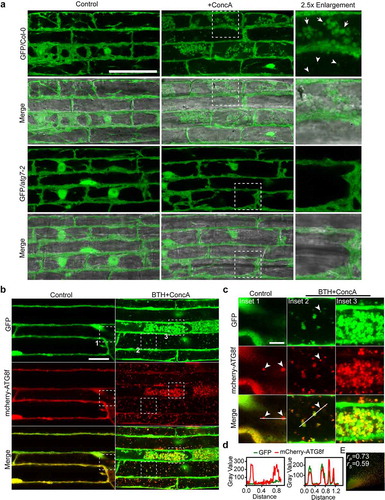
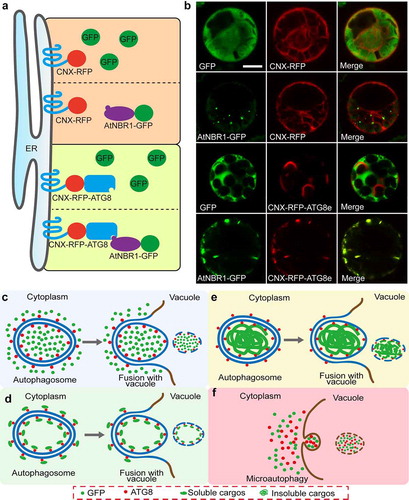
Figure 2. Proposed model for the nonselective cytoplasmic soluble protein and the selective cargos engulfed by autophagosomes
(A) in vivo recruitment model to identify cargos/receptors that degraded by selective autophagy. (B) The free GFP cannot be recruited to ER-localized CNX-RFP-ATG8e. The paired constructs were transiently co-expressed in Arabidopsis mesophyll protoplasts. AtNBR1-GFP was used as a positive control.Citation7 Bar = 10 μm. (C-F) Different pathways that may contribute to cytosol proteins accumulated and colocalized with ATG8 in the vacuole. The autophagosomes are formed to sequestrate partial cellular components containing cytosolic soluble proteins, like GFP, and subsequently fused with the vacuole to deliver the inclusions into the vacuolar lumen (C). For the soluble cargos, they are specially recognized and recruited by ATG8 to the double membrane of autophagosome. In this case, ring-like structures of these cytosolic cargos, such as C53, sometimes can be observed in cytoplasm (D). For the protein aggregates, they are recognized and engulfed by autophagosomes. In cytoplasm, the aggregates are colocalized with the autophagic marker ATG8 in a punctate pattern. Both of nonselective and the selective cytosolic materials were showed colocalization after the autophagic bodies accumulated in the vacuole (E). The cytoplasmic components might be directly engulfed by microautophagy at the level of the tonoplast or of the membranes of the MVB (multivesicular body) and then enter into the vacuolar lumen by invagination and scission (F).
Supplemental material