Figures & data
Figure 1. Synthesis of copolymer of methacrylic acid and butylmethacrylate.
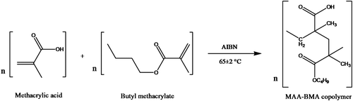
Figure 2. Percentage weight gain (swelling) of polymer membranes in buffers of increasing pH. Each bar represents mean ± SD (n = 3).
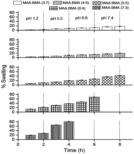
Figure 3. Percentage erosion of polymer membranes in buffers of increasing pH (− sign indicates weight loss). Each bar represents mean ± SD (n = 3).
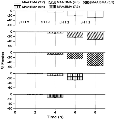
Figure 4. FTIR spectra of monomers (methacrylic acid and butyl methacrylate) and selected copolymers MAA:BMA (3:7 and 4:6).
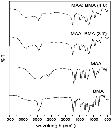
Figure 5. 1H NMR spectra of monomers (methacrylic acid and butyl methacrylate) and selected copolymers MAA:BMA (3:7 and 4:6).
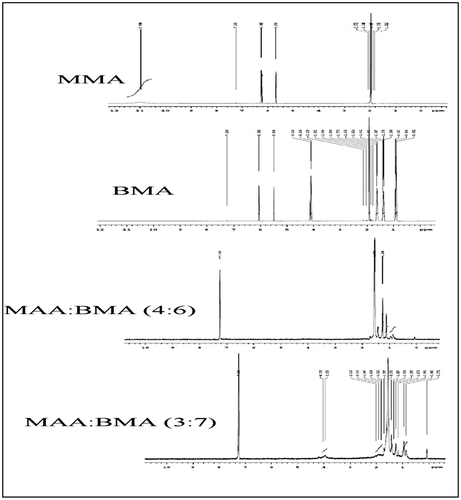
Table 1. Average molecular weights of copolymers MAA:BMA (3:7 and 4:6).
Figure 6. SEM images of microparticle formulation M3 (a) and (b); and M4 (c).

Table 2. Microspheres characterization.
Figure 7. Overlay showing PXRD diffractograms of aceclofenac, MAA:BMA (3:7 and 4:6), and formulation M3 and formulation M4.
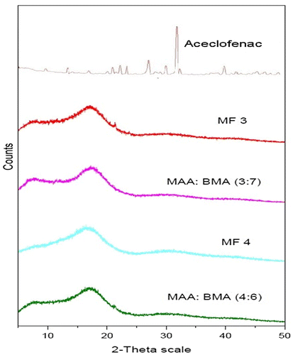
Figure 8. Overlay showing DSC thermograms of formulation M4 (A), copolymer MAA: BMA (4:6) (B), copolymer MAA:BMA (3:7) (C), formulation M3 (D), and aceclofenac (E).
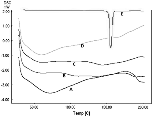
Figure 9. In vitro drug release profile of microparticle formulations M3 and M4. Each point represents mean ± SD (n = 3).
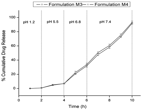
Table 3. Release kinetics of aceclofenac from microparticles using different kinetic equations.
Figure 10. Clinical activity scores during induction of colitis (from day 1 to day 9) and colon-to-body weight ratios (day 3, 5, 7 and 9) after TNBS administration. *Statistically significant (p < 0.05) compared with group I (healthy), as determined by one-way ANOVA followed by Dunnett’s test. Each bar represents the mean ± SD (n = 5).
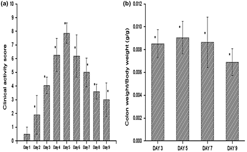
Figure 11. Clinical activity scores, colon-to-body weight ratio, and myeloperoxidase activity of group I (healthy group), group II (colitis group), group III (formulation treatment group), and group IV (standard treatment group). *Statistically significant (p < 0.05) compared with group II (colitis group), as determined by one-way ANOVA followed by Dunnett’s test. Each bar represents the mean ± SD (n = 5).
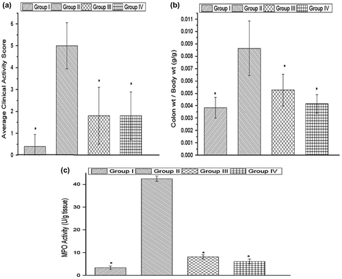
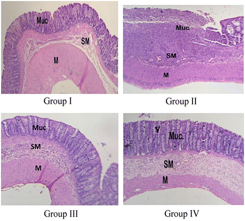
Figure 12. Microscopic image of the colon of rat belonging to group I (healthy group), group II (colitis group), group III (formulation treatment group), and group IV (standard treatment group).