Figures & data
Figure 1. The FTIR spectra of diphenyldithioperoxy anhydride, RAFT agent, and VEA monomer.
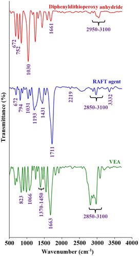
Figure 2. The 1H NMR spectra of the synthesized RAFT agent and VEA monomer (CDCl3, 25 °C).
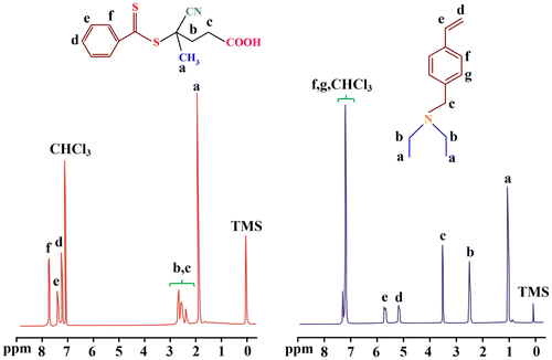
Figure 3. Absorbance spectra of diphenyldithioperoxy anhydride, and RAFT agent in dichloromethane solution.
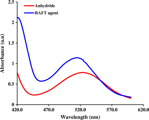
Figure 4. The FTIR spectra of the PHEMA, PHEMA-b-PVEA, and PSEMA-b-PVEA.
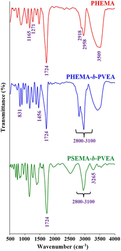
Figure 5. The 1H NMR spectra of the PHEMA and PHEMA-b-PVEA (DMSO-d6, 25 °C).
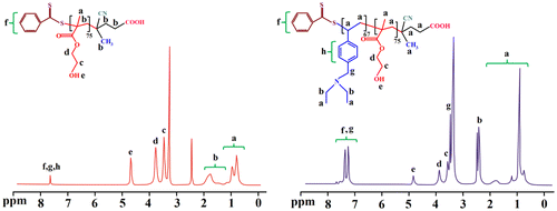
Figure 6. The 1H NMR spectra (D2O, 25 °C) for the PSEMA-b-PVEA ‘schizophrenic’ diblock copolymer recorded at pHs 3.0 and 10.0 using DCl and NaOD where appropriate.
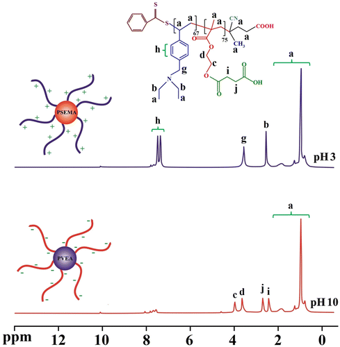
Figure 7. The DLS diagrams of the PSEMA-b-PVEA diblock copolymer at various pH values.
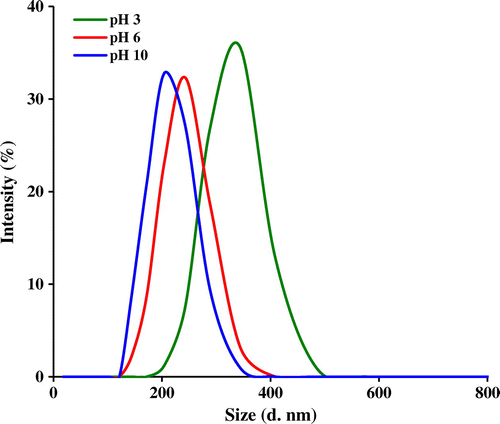
Figure 8. Variation of ξ potential with pH for a 0.50% (w/v) aqueous solution of the PSEMA-b-PVEA diblock copolymer.
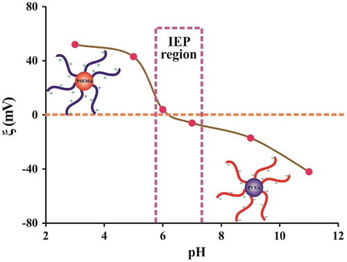
Figure 9. TEM micrographs of PSEMA-b-PVEA micelles at various pHs.
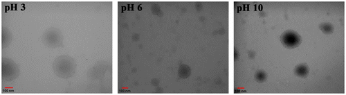
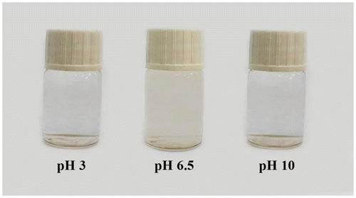
Figure 10. Digital photographs of PSEMA-b-PVEA diblock copolymer solution at pHs 3, 6.5, and 10.
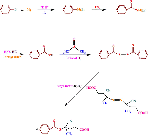
Scheme 1. Synthesis route of the RAFT agent.
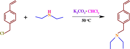
Scheme 2. Synthesis route of the VEA monomer.
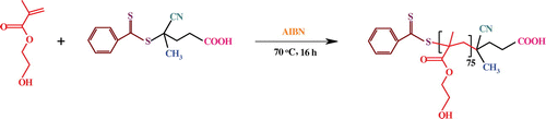
Scheme 3. Synthesis of PHEMA via RAFT polymerization.
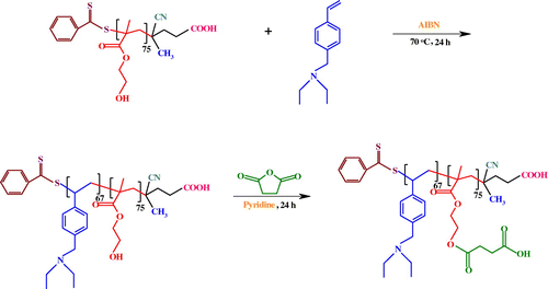
Scheme 4. Synthesis route of PHEMA-b-PVEA, and PSEMA-b-PVEA.
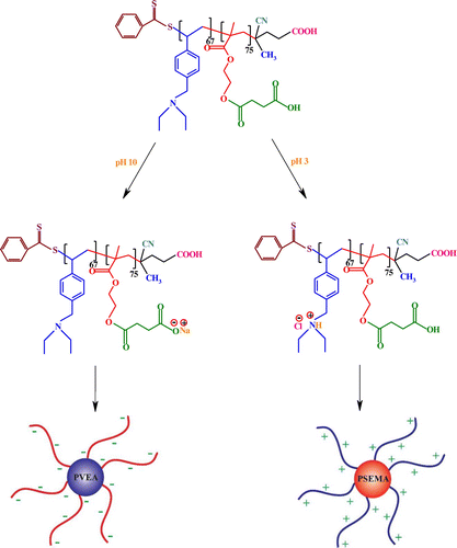
Scheme 5. The possible schematic structure of PSEMA-b-PVEA micelles at pHs 10.0 and 3.0.