Figures & data
Figure 1. Sequencing results of eight kinds of hemoglobin variants. The mutation sites were indicated by arrows. A: Hb Q-Thailand (HBA1:c.223G > C); B: Hb G-Chinese (HBA2:c.91G > C); C: Hb J-Broussais (HBA2:c.273G > T); D: Hb G-Taipei (HBB:c.68A > G); E: Hb Ottawa (HBA1:c.46G > C); F: Hb New York (HBB:c.341T > A); G: Hb G-Coushatta (HBB:c.68A > C); H: Hb J-Bangkok (HBB:c.170G > A).
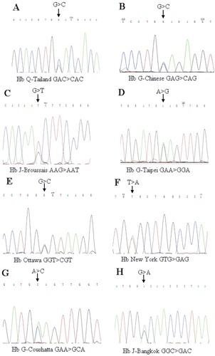
Table 1. The 45 cases of hemoglobin variants detected by electrophoresis and gene sequencing.
Figure 2. The historical migration waves of Hakka (Shaokwan was marked with ★ in the map).
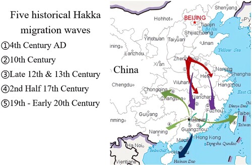
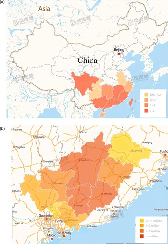
Figure 3. The distribution of Hakka population in Modern China. (a) The main inhabitant region of Hakka in southern China. (b) The distribution of Hakka in Shaokwan and its neighboring cities.