Figures & data
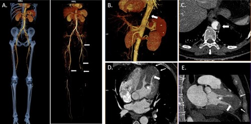
Figure 1. Three-dimensional reconstruction and computed tomography angiography (CTA) images of post-COVID-19 heparin-induced thrombocytopenia. (A) CTA three-dimensional image of multiple acute bilateral arterial thrombi (arrows) in the lower extremities suggesting thromboembolic origin with occlusion of the left popliteal artery. It is remarkable that there is no significant pre-existing aortic atherosclerosis. Three-dimensional reconstruction (B) and axial CTA (C) images showing a non-occlusive mural thrombus in the thoracoabdominal aortic hiatus. Four-chamber (D) and two-chamber views (E) revealing the presence of a large thrombus (65×32×34 mm) with a mobile distal pedicle (*) in the peripherally calcified apical left ventricular aneurysm (small arrows).