Figures & data
Figure 1. The results of the first serum protein electrophoresis and immunofixation electrophoresis (April 15, 2021). (A) shows that there was one M protein in the SPE. (B) shows that the immunotyping was IgG κ.
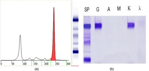
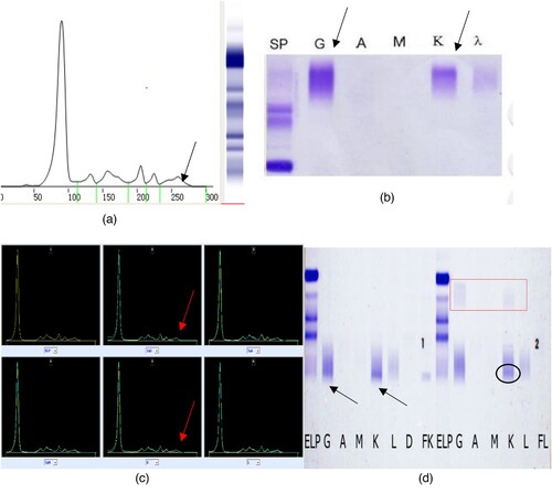
Figure 2. Results of the second serum protein electrophoresis, immunofixation electrophoresis, immunophenotyping and the Hydrashift 2/4 Daratumumab Assay (May 27, 2021). (A) (SPE) shows that there was one M proteins in the SPE, with an extra band (the arrow points). (B) (SIFE) shows that the immunotyping was IgG κ, with an extra band (the arrow points). (C) (immunophenotyping) also shows that the immunotyping was IgG κ, with an extra band (the arrow points). (D) (Hydrashift 2/4 Daratumumab Assay) shows the extra band was removed from the point of the arrow to the red box.
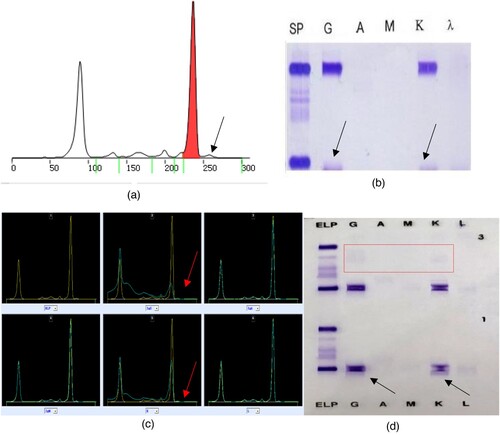
Figure 3. The results of the first serum protein electrophoresis and immunofixation electrophoresis (January 8, 2021). The serum protein electrophoresis in our hospital showed no M protein. Immunofixation electrophoresis + immunoglobulin light chain quantification: no obvious clonal bands were observed.
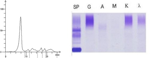
Figure 4. The results of the second serum protein electrophoresis and immunofixation electrophoresis (March 10, 2021). (A) shows that there was one M protein in the SPE. (B) shows that the report of the SIFE was IgG κ immunotyping.
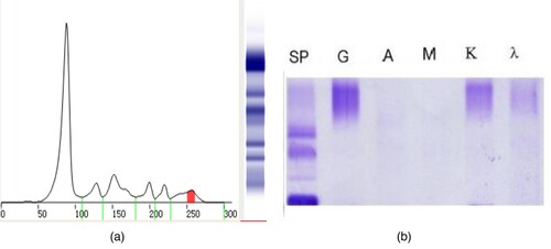
Figure 5. The results of serum protein electrophoresis, immunofixation electrophoresis, immunophenotyping and the Hydrashift 2/4 Daratumumab Assay (May 27, 2021). (A–C) shows that there maybe one M protein (the arrow points). (D) (Hydrashift 2/4 Daratumumab Assay) shows the ‘M’ protein was removed from the point of the arrow to the red box with an M protein (circled by an oval, with the immunophenotyping of κ light chain).