
Figures & data
Table 1. Demographic and clinicopathologic features of 306 patients receiving bendamustine treatment for lymphoid neoplasms.
Table 2. Demographic and clinicopathologic features of 58 patients with cytomegalovirus virologic reactivation and disease during bendamustine treatment for lymphoid neoplasms.
Figure 1. Infective complications of bendamustine-treated patients. (A). Patient 4 of the cytomegalovirus (CMV) disease cohort. Magnetic resonance imaging showed periventricular hyperintensities (arrows) indicative of ependymitis/ventriculitis. (B). Patient 5 of the CMV disease cohort. Upper endoscopy showing duodenal inflammation (left panel), and colonoscopy showing colitis (right panel). Histopathology of duodenal biopsy showed intranuclear CMV inclusion bodies, confirming CMV disease. (C). Patient 4 of the invasive fungal disease (IFD) cohort. Chest X ray showing bilateral consolidation. The patient was subsequently found to have cryptococcemia. (D). Patient 1 of the IFD cohort. Computed tomography (CT) scan of chest, showing wedge-shaped segmental lobar involvement (arrow) and consolidation (open arrow). Sputum grew Aspergillus niger. (E). Patient 3 of the IFD cohort. CT scan showed dense, well-circumscribed lesions (arrows). Bronchoalveolar aspirate grew Scedosporium apiospermum.
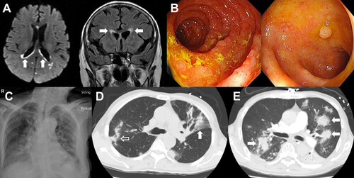
Table 3. Cytomegalovirus (CMV) disease in five patients treated with bendamustine.
Table 4. Invasive fungal diseases (IFD) in five patients treated with bendamustine.