
Figures & data
Table 1. Clinical features of the cancer patients with COVID-19.
Table 2. Assessment of the clinical and laboratory markers among the three patients’ groups.
Figure 1. Morphological characteristics of the peripheral blood cells in COVID-19 cancer patients showing: (A) Giant platelets. (B) Neutrophils with toxic granulations, bright mode. (C) Neutrophils with Pelger-Huet abnormality, bright mode. (D) Large sided aggressive looking monocytes with cytoplasmic vacuolations, 100× magnification.
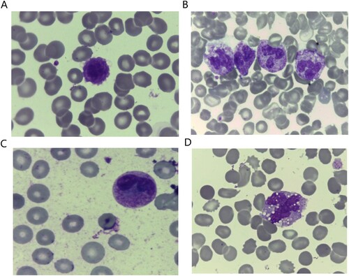
Figure 2. (A) Assessment of the Covicytes in patients’ groups. (B–E) Atypical lymphocytes with clumpy chromatin and basophilic cytoplasm with large granular tail (Covicytes), 100× magnification.
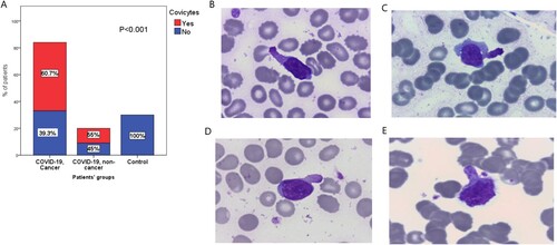
Figure 3. (A) The plasmacytoid lymphocyte, atypical lymphocyte with dark blue cytoplasm and slightly eccentric nucleus. (B) The monocytoid and ballerina looking lymphocytes were reactive lymphocytes that characteristically scalloping the neighboring cells, 100× magnification. (C) and (D) NK/T-cell lymphoma in leukaemic phase described by Leach M and Bain BJ, cited from the book ‘From the Image to the Diagnosis’, 1st edition. Page 58 [Citation25].
![Figure 3. (A) The plasmacytoid lymphocyte, atypical lymphocyte with dark blue cytoplasm and slightly eccentric nucleus. (B) The monocytoid and ballerina looking lymphocytes were reactive lymphocytes that characteristically scalloping the neighboring cells, 100× magnification. (C) and (D) NK/T-cell lymphoma in leukaemic phase described by Leach M and Bain BJ, cited from the book ‘From the Image to the Diagnosis’, 1st edition. Page 58 [Citation25].](/cms/asset/c4ee8707-c33f-444e-8d23-900f4c3b349d/yhem_a_2089830_f0003_oc.jpg)
Table 3. Association between the Covicytes and the clinical and laboratory features of the cancer COVID-19 patients.
Table 4. Association between Covicytes and clinical features of the non-cancer COVID-19 patients.
Data availability statement
All required data and materials are available upon request, except for the personal data of the patients.