Figures & data
Figure 1. Morphologic features of BM sample from the patient. (A) BM aspiration smear on admission in February 2020 revealed abnormal accumulations of immature cells containing multiple Auer rods in the cytoplasm (arrow) (100×). (B) BM aspiration smear in November 2020 after APL CR revealed erythroid cells were significantly increased, mainly composed of middle and late juvenile erythrocytes. (arrow) (100×). (C) BM biopsy on admission in February 2020 showing hypercellularity of immature atypical myeloid cells (H&E-stained image, 40×). (D) BM biopsy in November 2020 after APL CR. H&E-stained sections show active hyperplasia of nucleated myeloid and erythroid cells. The morphology of those cells was generally normal (40×).
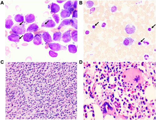
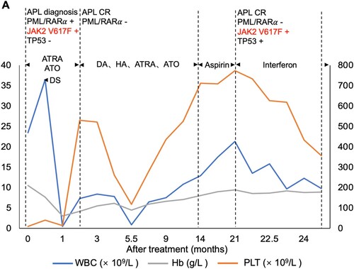
Figure 2. Clinical course of the patient. White blood counts (WBC), hemoglobin (Hb) and platelet (PLT) counts during the treatment. DS, differentiation syndrome; DA, daunorubicin + cytarabine; HA, harringtonine + cytarabine;