Figures & data
Table 1. 80% and 50% maximum inhibitory concentrations of different fractions.
Figure 1. Inhibition rates for MDA-MB-231 cells after treatment with isolated fractions of ‘Noble’ pomace. F1: mixture of flavonoids, phenolic acids and EA; F2: mixture of tannins; F3: mixture of anthocyanidins and EA.
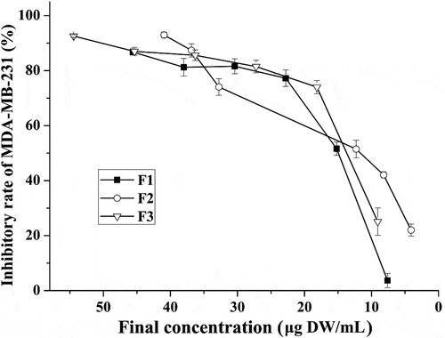
Table 2. Anti-oxidative activities of different fractions at low and high concentrations.
Figure 2. Plots from the cell cycle arrest assay using different fractions at low and high concentrations. Control: solvent treatment; F1 (F2, F3)_L: low concentrations of F1 (F2, F3); F1 (F2, F3)_H: high concentrations of F1 (F2, F3).
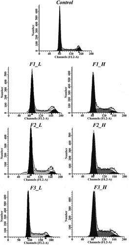
Table 3. Proportion of cells in different cell cycle phases after treatment with different fractions at high and low concentration (mean ± standard deviation).
Figure 3. Expression of cell cycle arrest proteins following treatment with different fractions at high or low concentrations. (a) Bands obtained from Western blot analysis; (b) bar charts of the mean integrated density of each band over the corresponding integrated density of GAPDH (control value adjusted to 1). ‘a, b, c’: the same letter above the bars indicates no significant differences (p ≥ 0.05). Control: solvent treatment; F1 (F2, F3)_L: low concentrations of F1 (F2, F3); F1 (F2, F3)_H: high concentrations of F1 (F2, F3).
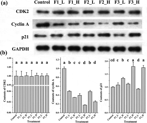
Table 4. Proportions of normal and apoptotic cells after treatment with different fractions at high and low concentrations (mean ± standard deviation).
Figure 4. Plots from the cell apoptosis assay for different fractions at low and high concentrations. Control: solvent treatment; F1 (F2, F3)_L: low concentrations of F1 (F2, F3); F1 (F2, F3)_H: high concentrations of F1 (F2, F3).
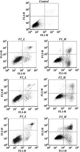
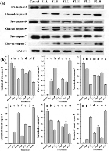
Figure 5. Contents of apoptosis proteins following treatment with different fractions at high or low concentrations. (a) Bands obtained from Western blotting; (b) bar charts of the mean integrated density of each band over the corresponding integrated density of GAPDH (control value adjusted to 1). ‘a, b, c’: the same letter above the bars indicates no significant differences (p ≥ 0.05). Control: solvent treatment; F1 (F2, F3)_L: low concentrations of F1 (F2, F3); F1 (F2, F3)_H: high concentrations of F1 (F2, F3).