Figures & data
Figure 1. Percentage of root length colonized by four individual fungi in wild-type ‘Gifu’ and in three symbiotic mutants of Lotus japonicus. Colonization in mutants was only by extraradical and intraradical hyphae. Bar indicates LSD at p<0.05.
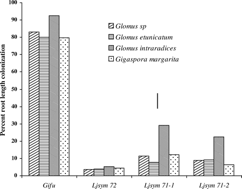
Table I Percentage root length colonized by external hyphae, appressoria, internal hyphae, arbuscules and vesicles of four different AM fungi.
Figure 2. Light microscopy of cleared and trypan blue stained roots of wild-type ‘Gifu’ (Figures A–D) and mutant Ljsym 72 (Figures E–H) of Lotus japonicus, colonized by Glomus sp. R-10 (A, E), Glomus etunicatum (B, F), Glomus intraradices (C, G) and Gigaspora margarita (D, H). eh, external hypahe, ap, appressoria, ih, internal hyphae, ar, arbuscules, v, vesicles. Bar represents 50 µm.
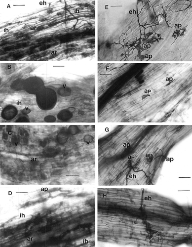
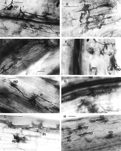
Figure 3. Light microscopy of cleared and trypan blue stained roots of mutant Ljsym 71-1 (Figures A–D) and mutant Ljsym 71-2 (Figures E–H) of Lotus japonicus, colonized by Glomus sp. R-10 (A, E), Glomus etunicatum (B, F), Glomus intraradices (C, G) and Gigaspora margarita (D, H). ap, appressoria, ih, internal hyphae. Bar represents 50 µm.