Figures & data
Figure 1. NTR activity in cotyledons (A) and embryonic axes (B) of P. sativum seeds during germination after imbibition with H2O or 5 mM Cd. Data are the mean of six independent measurements±SE. Each measurement was performed in an extract obtained from several germinating seeds.
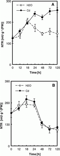
Figure 2. Western blot analysis of NTR expression in cotyledons (A) and embryonic axes (B) of P. sativum seeds during germination after imbibition with H2O or 5 mM Cd. Analysis was performed after SDS-PAGE of post mitochondrial proteins obtained from several germinating seeds (20 µg/track), transferred to nitrocellulose sheet and immunodetected with AtNTR antibody. The blot is representative of two experiments.
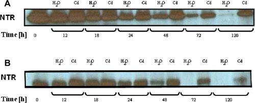
Figure 3. Western blot analysis of Trx expression in cotyledons (A,C) and embryonic axes (B,D) of P. sativum seeds during germination after imbibition with H2O or 5 mM Cd. Analysis was performed after SDS-PAGE of post mitochondrial proteins obtained from several germinating seeds (20 µg/track), transferred to nitrocellulose sheet and immunodetected with poplar Trx h3 and Trx h4 antibodies. The blot is representative of two experiments.
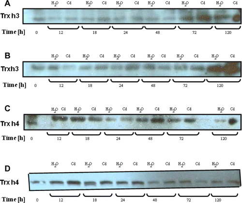
Figure 4. Trx activity in cotyledons (A) and embryonic axes (B) of P. sativum seeds during germination after imbibition with H2O or 5 mM Cd. Data are the mean of six independent measurements ±SE. Each measurement was performed in an extract obtained from several germinating seeds.
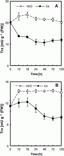
Figure 5. Effect of Cd ions on Trx activity in vitro. Poplar proteins (5 µg) were mixed with 5 µg Cd or equal volume of H2O (control) and incubated for different times before the assay which was performed using either the DTNB-NTR test (Trx h3) or the NADP-malate dehydrogenase activation test (Trx f). Data are the mean of six independent measurements ±SE.
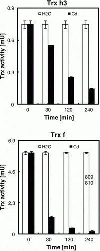
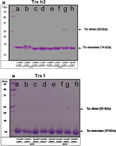
Figure 6. Effect of Cd binding on Trx. Poplar proteins (5 µg per lane) were mixed with 5 µg Cd, 30 mM β-MET and DTT (+) or equal volume of H2O (−) and incubated for 240 min before being subjected to 15% SDS-PAGE (a, Trx+ β-MET- DTT- Cd; b, Trx- β-MET- DTT- Cd; c, Trx+ β-MET+ DTT- Cd; d, Trx- β-MET+ DTT+ Cd; e, Trx + β-MET+ DTT+ Cd; f, Trx- β-MET+ DTT+ Cd; g, Trx- β-MET- DTT+ Cd; h, Trx + β-MET- DTT+ Cd). M – molecular weight markers are indicated in KDa. Trx dimer and mononer were stained with Coomassie bleue. Experiments were performed in duplicate.