
Figures & data
Table 1. Typical characteristics of the most common molecular imaging modalities for surgical guidance.
Table 2. Most common surgical applications of molecular imaging, summarizing surgical targets, the anatomies and molecular targets.
Figure 1. Decision making stages in a typical intraoperative molecular imaging surgery workflow and how these relate to red- (stop), orange- (slow down and approach with caution), and green- (go) light image guidance.
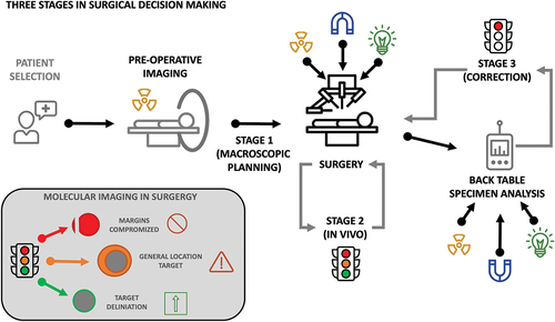
Figure 2. Examples of molecular image guidance technologies at different stages of the procedure. Stage 1, showing PET imaging (top and middle) and SPECT imaging (bottom) for planning of the intervention; Stage 2, showing freehand SPECT navigation (top), DROP-IN gamma probe and gamma camera guidance (middle) and fluorescence flow analysis [Citation38] and optical guidance, for in vivo guidance during the intervention; Stage 3, showing DROP-IN beta probe analysis (top) and PET specimen imager [Citation39] (bottom), for ex vivo back-table analysis during the intervention. Non referenced images originate from authors personal collection. No changes have been made to the referenced images, which are licensed under CC by 4.0 (https://creativecommons.org/licenses/by/4.0/).
![Figure 2. Examples of molecular image guidance technologies at different stages of the procedure. Stage 1, showing PET imaging (top and middle) and SPECT imaging (bottom) for planning of the intervention; Stage 2, showing freehand SPECT navigation (top), DROP-IN gamma probe and gamma camera guidance (middle) and fluorescence flow analysis [Citation38] and optical guidance, for in vivo guidance during the intervention; Stage 3, showing DROP-IN beta probe analysis (top) and PET specimen imager [Citation39] (bottom), for ex vivo back-table analysis during the intervention. Non referenced images originate from authors personal collection. No changes have been made to the referenced images, which are licensed under CC by 4.0 (https://creativecommons.org/licenses/by/4.0/).](/cms/asset/7c43a6d6-44ac-4fab-b88d-61f49270536c/ierd_a_2341102_f0002_oc.jpg)