Figures & data
Figure 1. Virtual pelvic radiographs were made using computer simulation. The virtual pelvis was represented by 5 points: the most caudal part of the two teardrops, the most distal part of the two sacro-iliac joints, and the midpoint of the most cranial part of the pelvic symphysis. The virtual total hip replacement was represented by the center of the acetabular cup (marked with a cross-hair) and the center of the femoral head (marked with a circle). The projected distance between these two represents both polyethylene wear and the projected wear direction. Virtual pelvic rotations were performed around the x- and the z-axes, and wear directions were altered in the coronal plane and the sagittal plane. The effects on the virtual wear measurements were then quantified.
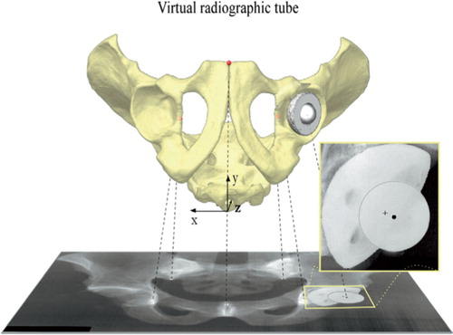
Figure 2. The correlation between the orientation of the virtual pelvis and the degree of underestimation of wear. The ranges of pelvic orientation were initially selected arbitrarily to be larger than those found in clinical settings. The yellow area represents variations in pelvic orientation found in clinical material, and a maximum of 2.6% of underestimation of wear was found.
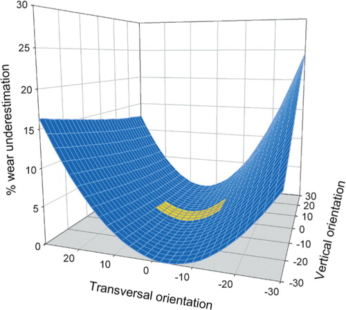
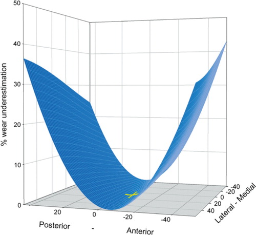
Figure 3. A maximum of 42% underestimation of wear was found when the wear directions were altered in the coronal plane (lateral-medial) and the sagittal plane (anteriorposterior) within ranges found clinically. The yellow crosshair marks the most common direction of wear described by Yamaguchi et al. (date) (8.1 degrees laterally and 4.1 degrees anteriorly). Variations in direction of wear in the coronal plane hade little effect on wear measurements since the parallelism between the direction of wear and the film plane remains unaltered.