Figures & data
Table 1. Sodium channel distribution and classification based on sensitivity to tetrodotoxin (TTX) block
Figure 1. NaV channel structural topology, highlighting common ligand binding sites and significant structural features. Domains D1-D4 are represented in different colors while β subunits are shown in gray.
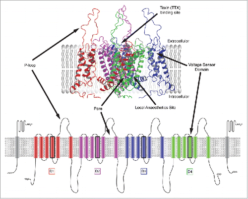
Figure 2. Sodium channel states: NaV channels cycle between 3 states: open, closed (resting) and inactivated. They cycle from the closed (resting) state to the open state upon membrane depolarisation. The channel is open for less than a millisecond prior to inactivation. The inactivated state reprimes to the resting state when the cell membrane potential has returned to a hyperpolarised resting potential. Sodium channel current: The current associated with the cycling of sodium channels through the resting, open and inactivated state.
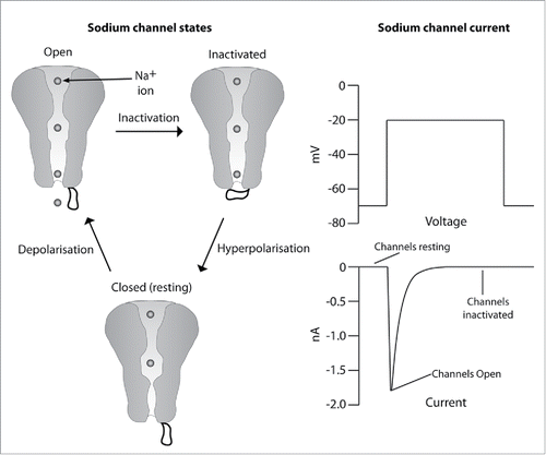
Figure 3. Selected toxin modulators.
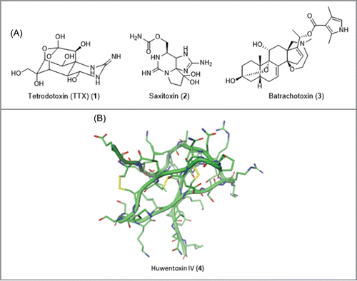
Figure 4. Selected first generation sodium channel modulator drugs.
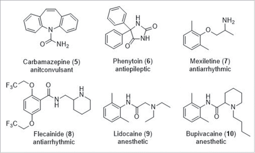
Figure 5. Early examples of second generation sodium channel modulators.
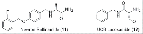
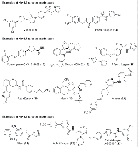
Figure 6. Examples of NaV subtype selective targeting modulators.