7,137
Views
66
CrossRef citations to date
0
Altmetric
Report

Pharmacokinetic properties of IgG and various Fc fusion proteins in mice

, , , , , & show all
Pages 120-128 | Received 03 Sep 2015, Accepted 21 Oct 2015, Published online: 30 Dec 2015

Figures & data

Figure 1. Schematic composition of the various proteins. Carbohydrates are shown as hexagonal gray symbols.
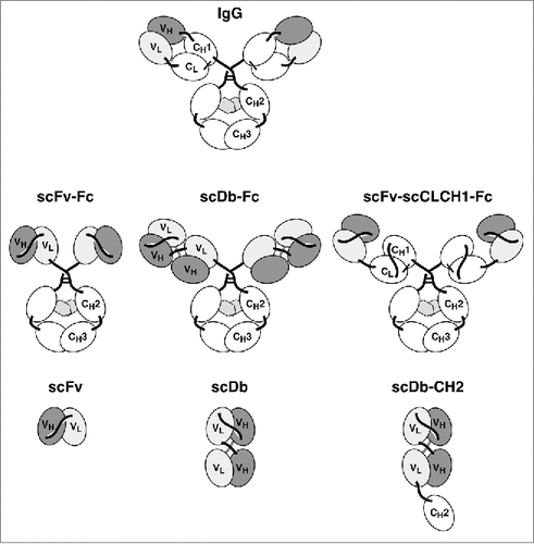
Figure 1. Schematic composition of the various proteins. Carbohydrates are shown as hexagonal gray symbols.

Figure 2. Pharmacokinetics of IgG1 and scFv-Fc fusion proteins. a) Anti-CEA antibody molecules. b) Anti-EGFR antibody molecules. CD1 mice received a single injection of 25 µg protein and serum concentrations were determined by ELISA.
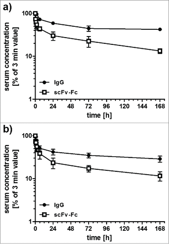
Figure 2. Pharmacokinetics of IgG1 and scFv-Fc fusion proteins. a) Anti-CEA antibody molecules. b) Anti-EGFR antibody molecules. CD1 mice received a single injection of 25 µg protein and serum concentrations were determined by ELISA.

Table 1 Biochemical and pharmacokinetic properties

Figure 3. a) SDS-PAGE analysis of IgG and Fc fusion proteins (lanes 1 and 7, IgG1 produced in CHO cells; lanes 2 and 8, scFv-Fc; lanes 3 and 9, scDb-Fc; lanes 4 and 10, scFv-scCLCH1-Fc; lanes 5 and 11, IgG1 produced in HEK293 cells; lanes 6 and 12, scDb-CH2) analyzed under non-reducing (1–6) or reducing (7–12) conditions. Proteins were stained with Coomassie blue. b) Size-exclusion chromatography of IgG and Fc fusion proteins as indicated.
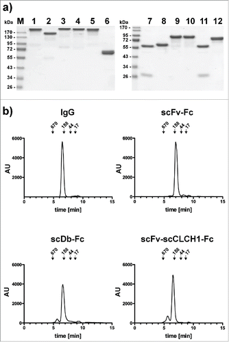
Figure 3. a) SDS-PAGE analysis of IgG and Fc fusion proteins (lanes 1 and 7, IgG1 produced in CHO cells; lanes 2 and 8, scFv-Fc; lanes 3 and 9, scDb-Fc; lanes 4 and 10, scFv-scCLCH1-Fc; lanes 5 and 11, IgG1 produced in HEK293 cells; lanes 6 and 12, scDb-CH2) analyzed under non-reducing (1–6) or reducing (7–12) conditions. Proteins were stained with Coomassie blue. b) Size-exclusion chromatography of IgG and Fc fusion proteins as indicated.

Figure 4. ELISA of binding of IgG and Fc fusion proteins to immobilized CEA. Bound proteins were detected with an anti-Fc HRP-conjugated antibody.
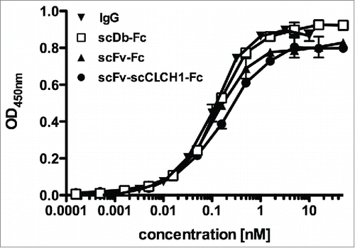
Figure 4. ELISA of binding of IgG and Fc fusion proteins to immobilized CEA. Bound proteins were detected with an anti-Fc HRP-conjugated antibody.

Figure 5. a) Thermal melting points of IgG and Fc fusion proteins determined by dynamic light scattering using 1°C intervals and 2 min equilibration time. A drastic increase in mean count rates is defined as melting point and indicated by dashed lines. b) In vitro serum stability of IgG and Fc fusion proteins. Proteins were incubated with mouse serum at 37°C for up to 7 d and active protein was determined by ELISA (IgG and Fc fusion proteins with an anti-Fc antibody, scDb-CH2 either with an anti-His-tag antibody or protein L).
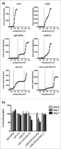
Figure 5. a) Thermal melting points of IgG and Fc fusion proteins determined by dynamic light scattering using 1°C intervals and 2 min equilibration time. A drastic increase in mean count rates is defined as melting point and indicated by dashed lines. b) In vitro serum stability of IgG and Fc fusion proteins. Proteins were incubated with mouse serum at 37°C for up to 7 d and active protein was determined by ELISA (IgG and Fc fusion proteins with an anti-Fc antibody, scDb-CH2 either with an anti-His-tag antibody or protein L).

Figure 6. a) QCM measurements of binding of IgG and Fc fusion proteins to immobilized mouse FcRn (at a density of 50 Hz) at pH 6.0 or pH 7.4. For comparison, binding curves are shown for 100 nM protein, except for scDb-CH2 which was analyzed at 1 µM. b) Analysis of dissociation of proteins bound to mouse FcRn (at pH 6.0) by shifting pH to 7.4. c) Analysis of dissociation of IgG and scFv-Fc fusion protein to human FcRn at pH 6.0 by shifting pH to 7.4.
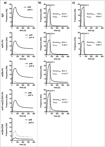
Figure 6. a) QCM measurements of binding of IgG and Fc fusion proteins to immobilized mouse FcRn (at a density of 50 Hz) at pH 6.0 or pH 7.4. For comparison, binding curves are shown for 100 nM protein, except for scDb-CH2 which was analyzed at 1 µM. b) Analysis of dissociation of proteins bound to mouse FcRn (at pH 6.0) by shifting pH to 7.4. c) Analysis of dissociation of IgG and scFv-Fc fusion protein to human FcRn at pH 6.0 by shifting pH to 7.4.

Table 2 Binding to mouse FcRn

Figure 7. a) Pharmacokinetics of IgG and Fc fusion proteins. b) Pharmacokinetics of scFv as well as scDb and scDb-CH2. CD1 mice received a single injection of 25 µg protein and serum concentrations were determined by ELISA. c) Statistical analysis of terminal half-lives and AUCs of the IgGs and Fc fusion proteins.
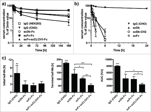
Figure 7. a) Pharmacokinetics of IgG and Fc fusion proteins. b) Pharmacokinetics of scFv as well as scDb and scDb-CH2. CD1 mice received a single injection of 25 µg protein and serum concentrations were determined by ELISA. c) Statistical analysis of terminal half-lives and AUCs of the IgGs and Fc fusion proteins.

Reprints and Corporate Permissions

Please note: Selecting permissions does not provide access to the full text of the article, please see our help page How do I view content?

To request a reprint or corporate permissions for this article, please click on the relevant link below:

Academic Permissions

Please note: Selecting permissions does not provide access to the full text of the article, please see our help page How do I view content?

Obtain permissions instantly via Rightslink by clicking on the button below:

If you are unable to obtain permissions via Rightslink, please complete and submit this Permissions form. For more information, please visit our Permissions help page.